Wei Xuan, Lv Han, Chen Qian, Wang Zhaodi, Liu Chunli, Zhao Pengfei, Gong Shusheng, Yang Zhenghan, Wang Zhenchang
Department of Radiology, Beijing Friendship Hospital, Capital Medical University, Beijing, China.
Department of Otolaryngology Head and Neck Surgery, Beijing Friendship Hospital, Capital Medical University, Beijing, China.
Front Neurosci. 2021 Mar 5;15:633364. doi: 10.3389/fnins.2021.633364. eCollection 2021.
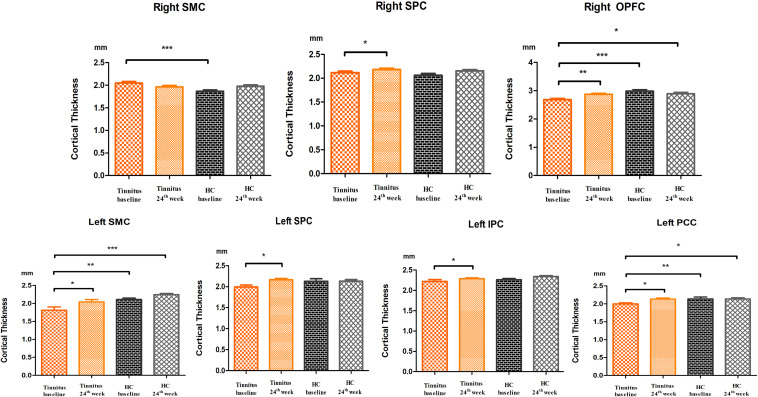
This study aimed to explore brain surface-based morphometry cortical thickness changes in patients with idiopathic tinnitus before and after 24 weeks of sound therapy. In this prospective observational study, we recruited 33 tinnitus patients who had undergone 24 weeks of sound therapy and 26 matched healthy controls. For the two groups of subjects, a 3D-BRAVO pulse sequence was acquired both at baseline and at the 24th week. Structural image data preprocessing was performed using the DPABISurf toolbox. The Tinnitus Handicap Inventory (THI) score was assessed to determine the severity of tinnitus before and after treatment. Two-way mixed-model analysis of variance (ANOVA) and Pearson's correlation analysis were used in the statistical analysis. Student-Newman-Keuls (SNK) tests were used in the analysis. Significantly lower cortical thickness was found in the left somatosensory and motor cortex (SMC), left posterior cingulate cortex (PCC), and right orbital and polar frontal cortex (OPFC) of the participants in the tinnitus group at baseline than in the participants in the HC group at baseline and after 24 weeks; in the tinnitus group, significantly higher cortical thickness was found after the 24 weeks sound therapy in comparison to the baseline in the left SMC, bilateral superior parietal cortex (SPC), left inferior parietal cortex (IPC), left PCC, and right OPFC. In the HC group, no statistically significant difference in cortical thickness was found after the 24 weeks treatment in comparison to the baseline in the bilateral SMC, bilateral SPC, left IPC, left PCC, or right OPFC. The changes in cortical thickness before and after sound therapy can provide certain reference values for clinical tinnitus treatment. These brain regions could serve as potential targets for neuroimaging.
本研究旨在探讨特发性耳鸣患者在接受24周声治疗前后基于脑表面形态测量的皮质厚度变化。在这项前瞻性观察研究中,我们招募了33名接受了24周声治疗的耳鸣患者和26名匹配的健康对照者。对于两组受试者,在基线和第24周均采集了3D - BRAVO脉冲序列。使用DPABISurf工具箱进行结构图像数据预处理。评估耳鸣障碍量表(THI)得分以确定治疗前后耳鸣的严重程度。统计分析采用双向混合模型方差分析(ANOVA)和Pearson相关分析。分析中使用了Student - Newman - Keuls(SNK)检验。在基线时,耳鸣组参与者的左侧躯体感觉和运动皮层(SMC)、左侧后扣带回皮层(PCC)以及右侧眶额和极前额皮层(OPFC)的皮质厚度显著低于健康对照组参与者在基线时和24周后的皮质厚度;在耳鸣组中,与基线相比,24周声治疗后左侧SMC、双侧顶上叶皮层(SPC)、左侧顶下叶皮层(IPC)、左侧PCC和右侧OPFC的皮质厚度显著增加。在健康对照组中,与基线相比,24周治疗后双侧SMC、双侧SPC、左侧IPC、左侧PCC或右侧OPFC的皮质厚度无统计学显著差异。声治疗前后皮质厚度的变化可为临床耳鸣治疗提供一定的参考价值。这些脑区可作为神经影像学的潜在靶点。