• 文献检索
  • 文档翻译
  • 深度研究
  • 学术资讯
  • Suppr Zotero 插件Zotero 插件
  • 邀请有礼
  • 套餐&价格
  • 历史记录
应用&插件
Suppr Zotero 插件Zotero 插件浏览器插件Mac 客户端Windows 客户端微信小程序
定价
高级版会员购买积分包购买API积分包
服务
文献检索文档翻译深度研究API 文档MCP 服务
关于我们
关于 Suppr公司介绍联系我们用户协议隐私条款
关注我们

Suppr 超能文献

核心技术专利:CN118964589B侵权必究
粤ICP备2023148730 号-1Suppr @ 2026

文献检索

告别复杂PubMed语法,用中文像聊天一样搜索,搜遍4000万医学文献。AI智能推荐,让科研检索更轻松。

立即免费搜索

文件翻译

保留排版,准确专业,支持PDF/Word/PPT等文件格式,支持 12+语言互译。

免费翻译文档

深度研究

AI帮你快速写综述,25分钟生成高质量综述,智能提取关键信息,辅助科研写作。

立即免费体验

通过追踪整个结节的剪切波弹性,发现甲状腺癌的弹性具有很高的可重复性和与纤维化的一致性。

Shear wave elasticity by tracing total nodule showed high reproducibility and concordance with fibrosis in thyroid cancer.

机构信息

Division of Endocrinology and Metabolism, Department of Internal Medicine, Soonchunhyang University Hospital, Soonchunhyang University College of Medicine, 59 Daesagwan-ro, Yongsan-gu, Seoul, 140-743, Republic of Korea.

Elim Thyroid Clinic, Seoul, South Korea.

出版信息

BMC Cancer. 2020 Feb 12;20(1):118. doi: 10.1186/s12885-019-6437-z.

DOI:10.1186/s12885-019-6437-z
PMID:32050941
原文链接:https://pmc.ncbi.nlm.nih.gov/articles/PMC7014777/
Abstract

BACKGROUND

Although shear wave elastography (SWE) is reported to be useful in detecting malignant thyroid nodules, it shows a wide range of cut-off values of elasticity index (EI) in detecting malignant nodules. The cause of discrepancy remains unclear. Fibrosis of the tumors is known to increase the EI in SWE, and matching of SWE and surgical histopathology has not been fully illustrated in thyroid cancer. We aimed to evaluate the reproducibility of the new total nodular region of interest (ROI) method excluding the subjective features of focal circular ROI placement and to determine the lesion that causes the elevation of EI on SWE and on histopathology.

METHODS

A total of 29 thyroid cancers from 28 patients were included. We evaluated the reproducibility of EI in the new total nodular ROI using Q-Box Trace program and compared the EI in focal nodular ROI using a 3-mm circular area. We analyzed the correlation between fibrosis and EI.

RESULT

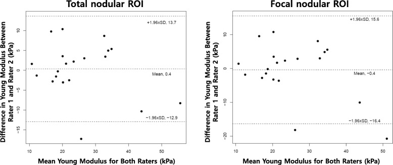
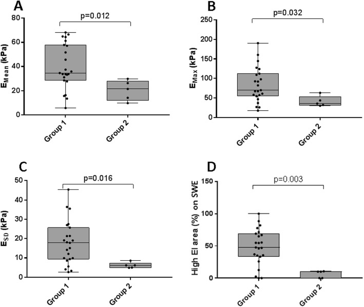
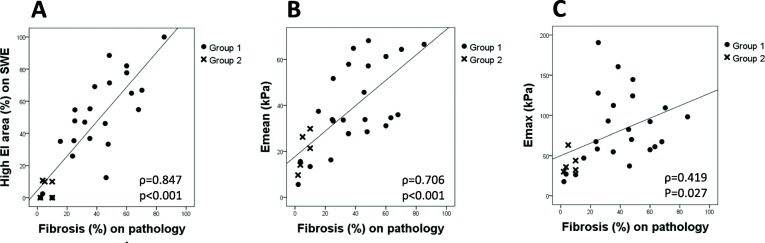
The coefficient of variation (CV) of the intrarater assay was significantly lower in total nodular ROI than in focal nodular ROI within the image in rater 1 (1.7% vs. 13.4%, p < 0.001) and in rater 2 (1.4% vs. 16.9%, p < 0.001) and in different images in rater 1 (7.6% vs. 12.3%, p = 0.040) and in rater 2 (7.5% vs. 19.8%, p = 0.004). Moreover, CV of the interrater assay showed similar results (14.9% vs. 19%, p = 0.030). Interrater intraclass correlation coefficient showed better agreement in total nodular ROI than in focal nodular ROI (0.863 vs. 0.783). The degree of fibrosis on histopathology showed significant correlations with EI (E, p < 0.001; E, p = 0.027), and the location of fibrosis was concordant with the high EI area on SWE.

CONCLUSION

Our study revealed that the new total nodular ROI method showed higher reproducibility and better agreement in intra- and interrater assay than the focal nodular ROI method, suggesting a valuable and standardized method in clinical practice. Moreover, our results showed that fibrosis in the histopathology increased EI on SWE and might lead to the discrepancy of the cut-off values in detecting thyroid cancer.

摘要

背景

尽管剪切波弹性成像(SWE)已被证实可用于检测甲状腺恶性结节,但在检测恶性结节时,其弹性指数(EI)的截断值范围较宽。造成这种差异的原因尚不清楚。肿瘤纤维化已知会增加 SWE 中的 EI,并且 SWE 与手术组织病理学的匹配尚未在甲状腺癌中得到充分说明。我们旨在评估新的全结节感兴趣区域(ROI)方法排除焦点圆形 ROI 放置的主观特征的可重复性,并确定在 SWE 和组织病理学上导致 EI 升高的病变。

方法

共纳入 28 例患者的 29 例甲状腺癌。我们使用 Q-Box Trace 程序评估了新的全结节 ROI 中 EI 的可重复性,并比较了使用 3mm 圆形区域的焦点结节 ROI 中的 EI。我们分析了纤维化与 EI 的相关性。

结果

在评分者 1 (1.7%对 13.4%,p<0.001)和评分者 2 (1.4%对 16.9%,p<0.001)的图像内,以及评分者 1 (7.6%对 12.3%,p=0.040)和评分者 2 (7.5%对 19.8%,p=0.004)的不同图像内,全结节 ROI 的组内变异系数(CV)明显低于焦点结节 ROI。此外,组间 CV 也显示出相似的结果(14.9%对 19%,p=0.030)。组间内评分者的组内相关系数显示,全结节 ROI 比焦点结节 ROI 具有更好的一致性(0.863 对 0.783)。组织病理学上纤维化的程度与 EI 显著相关(E,p<0.001;E,p=0.027),纤维化的位置与 SWE 上高 EI 区域一致。

结论

本研究表明,与焦点结节 ROI 方法相比,新的全结节 ROI 方法在组内和组间评估中具有更高的可重复性和更好的一致性,提示在临床实践中具有重要价值和标准化方法。此外,我们的研究结果表明,组织病理学上的纤维化增加了 SWE 上的 EI,可能导致在检测甲状腺癌时的截断值差异。

https://cdn.ncbi.nlm.nih.gov/pmc/blobs/3803/7014777/68f90cdcac01/12885_2019_6437_Fig4_HTML.jpg
https://cdn.ncbi.nlm.nih.gov/pmc/blobs/3803/7014777/b92d222aa3b3/12885_2019_6437_Fig1_HTML.jpg
https://cdn.ncbi.nlm.nih.gov/pmc/blobs/3803/7014777/880d0a377358/12885_2019_6437_Fig2_HTML.jpg
https://cdn.ncbi.nlm.nih.gov/pmc/blobs/3803/7014777/1a35894b53d1/12885_2019_6437_Fig3_HTML.jpg
https://cdn.ncbi.nlm.nih.gov/pmc/blobs/3803/7014777/68f90cdcac01/12885_2019_6437_Fig4_HTML.jpg

相似文献

1
Shear wave elasticity by tracing total nodule showed high reproducibility and concordance with fibrosis in thyroid cancer.通过追踪整个结节的剪切波弹性,发现甲状腺癌的弹性具有很高的可重复性和与纤维化的一致性。
BMC Cancer. 2020 Feb 12;20(1):118. doi: 10.1186/s12885-019-6437-z.
2
Is the reproducibility of shear wave elastography of thyroid nodules high enough for clinical use? A methodological study.甲状腺结节剪切波弹性成像的可重复性是否足以用于临床?一项方法学研究。
Clin Endocrinol (Oxf). 2017 Apr;86(4):606-613. doi: 10.1111/cen.13295. Epub 2017 Jan 16.
3
Shear wave elastography of thyroid nodules in routine clinical practice: preliminary observations and utility for detecting malignancy.常规临床实践中的甲状腺结节剪切波弹性成像:初步观察及其在检测恶性肿瘤中的应用。
Eur Radiol. 2012 Nov;22(11):2397-406. doi: 10.1007/s00330-012-2495-1. Epub 2012 May 30.
4
Reappraisal of shear wave elastography as a diagnostic tool for identifying thyroid carcinoma.重新评估剪切波弹性成像作为诊断甲状腺癌的工具。
Endocr Connect. 2019 Aug;8(8):1195-1205. doi: 10.1530/EC-19-0324.
5
Utility of shear wave elastography to detect papillary thyroid carcinoma in thyroid nodules: efficacy of the standard deviation elasticity.剪切波弹性成像技术在甲状腺结节中检测甲状腺乳头状癌的效用:标准差弹性的效能。
Korean J Intern Med. 2019 Jul;34(4):850-857. doi: 10.3904/kjim.2016.326. Epub 2018 Feb 23.
6
Shear wave elastography of thyroid nodules for the prediction of malignancy in a large scale study.在一项大规模研究中,甲状腺结节的剪切波弹性成像用于预测恶性肿瘤。
Eur J Radiol. 2015 Mar;84(3):407-412. doi: 10.1016/j.ejrad.2014.11.019. Epub 2014 Nov 27.
7
Shear wave elastography in the diagnosis of thyroid nodules: feasibility in the case of coexistent chronic autoimmune Hashimoto's thyroiditis.剪切波弹性成像在甲状腺结节诊断中的应用:合并慢性自身免疫性桥本甲状腺炎的可行性。
Clin Endocrinol (Oxf). 2012 Jan;76(1):137-41. doi: 10.1111/j.1365-2265.2011.04170.x.
8
Shear wave elastography combined with the thyroid imaging reporting and data system for malignancy risk stratification in thyroid nodules.剪切波弹性成像联合甲状腺影像报告和数据系统用于甲状腺结节恶性风险分层
Oncotarget. 2017 Jun 27;8(26):43406-43416. doi: 10.18632/oncotarget.15018.
9
The influence of precompression on elasticity of thyroid nodules estimated by ultrasound shear wave elastography.超声剪切波弹性成像技术测量甲状腺结节的预压缩对其弹性的影响。
Eur Radiol. 2016 Aug;26(8):2845-52. doi: 10.1007/s00330-015-4108-2. Epub 2015 Nov 25.
10
Comparison of strain elastography, point shear wave elastography using acoustic radiation force impulse imaging and 2D-shear wave elastography for the differentiation of thyroid nodules.应变弹性成像、声辐射力脉冲成像点剪切波弹性成像与二维剪切波弹性成像在甲状腺结节鉴别诊断中的比较。
PLoS One. 2018 Sep 17;13(9):e0204095. doi: 10.1371/journal.pone.0204095. eCollection 2018.

引用本文的文献

1
Assessment of Ultrasound-Controlled Diagnostic Methods for Thyroid Lesions and Their Associated Costs in a Tertiary University Hospital in Spain.西班牙一家三级大学医院中甲状腺病变的超声控制诊断方法及其相关成本评估
J Clin Med. 2025 Aug 6;14(15):5551. doi: 10.3390/jcm14155551.
2
Analysis of risk factors and application of risk management strategies in hemodialysis patients complicated with heart failure.血液透析合并心力衰竭患者的危险因素分析及风险管理策略的应用
Front Cardiovasc Med. 2025 Jun 16;12:1600223. doi: 10.3389/fcvm.2025.1600223. eCollection 2025.
3
Ultrasound Assessment of the Gastrocnemius Muscle as a Potential Tool for Identifying Sarcopenia in Patients with Type 2 Diabetes.

本文引用的文献

1
Differentiation of Benign and Malignant Thyroid Nodules by Using Comb-push Ultrasound Shear Elastography: A Preliminary Two-plane View Study.应用梳状推扫超声剪切弹性成像技术鉴别甲状腺良恶性结节:初步的双平面研究。
Acad Radiol. 2018 Nov;25(11):1388-1397. doi: 10.1016/j.acra.2018.02.025. Epub 2018 Mar 21.
2
The Preciseness in Diagnosing Thyroid Malignant Nodules Using Shear-Wave Elastography.使用剪切波弹性成像诊断甲状腺恶性结节的精确性。
Med Sci Monit. 2018 Feb 2;24:671-677. doi: 10.12659/msm.904703.
3
Shear wave elastography and Afirma™ gene expression classifier in thyroid nodules with indeterminate cytology: a comparison study.
超声评估腓肠肌作为2型糖尿病患者肌肉减少症潜在诊断工具的研究
Diabetes Metab Syndr Obes. 2023 Oct 30;16:3435-3444. doi: 10.2147/DMSO.S435517. eCollection 2023.
4
Combined Shear Wave Elastography and EU TIRADS in Differentiating Malignant and Benign Thyroid Nodules.联合剪切波弹性成像与欧盟甲状腺影像报告和数据系统在鉴别甲状腺良恶性结节中的应用
Cancers (Basel). 2022 Nov 10;14(22):5521. doi: 10.3390/cancers14225521.
5
Efficacy of Differential Diagnosis of Thyroid Nodules by Shear Wave Elastography-the Stiffness Map.剪切波弹性成像-硬度图对甲状腺结节的鉴别诊断效能
J Endocr Soc. 2021 Oct 3;5(11):bvab154. doi: 10.1210/jendso/bvab154. eCollection 2021 Nov 1.
6
Novel High-Quality Sonographic Methods to Diagnose Muscle Wasting in Long-Stay Critically Ill Patients: Shear Wave Elastography, Superb Microvascular Imaging and Contrast-Enhanced Ultrasound.新型高质量超声诊断方法诊断长期重症患者肌肉减少症:剪切波弹性成像、超微血流成像和超声造影。
Nutrients. 2021 Jun 29;13(7):2224. doi: 10.3390/nu13072224.
7
Ultrasound Shear Wave Elastography and Transient Optical Coherence Elastography: Side-by-Side Comparison of Repeatability and Accuracy.超声剪切波弹性成像与瞬态光学相干弹性成像:重复性和准确性的并列比较
IEEE Open J Eng Med Biol. 2021;2:179-186. doi: 10.1109/ojemb.2021.3075569. Epub 2021 Apr 27.
8
Evaluation of thyroid nodules by shear wave elastography: a review of current knowledge.剪切波弹性成像评估甲状腺结节:当前知识综述。
J Endocrinol Invest. 2021 Oct;44(10):2043-2056. doi: 10.1007/s40618-021-01570-z. Epub 2021 Apr 16.
9
Decreased Size of Mammary Tumors Caused by Preoperative Treatment with Aglepristone in Female Domestic Dogs () Do Not Influence the Density of the Benign Neoplastic Tissue Measured Using Shear Wave Elastography Technique.孕畜酮术前治疗导致雌性家犬乳腺肿瘤体积减小()并不影响使用剪切波弹性成像技术测量的良性肿瘤组织密度。
Animals (Basel). 2021 Feb 18;11(2):527. doi: 10.3390/ani11020527.
剪切波弹性成像与 Afirma™ 基因表达分类器在甲状腺细胞学不确定结节中的对比研究。
Endocrine. 2018 Mar;59(3):573-584. doi: 10.1007/s12020-017-1509-9. Epub 2018 Jan 19.
4
The 2017 Bethesda System for Reporting Thyroid Cytopathology.2017 年甲状腺细胞病理学报告的贝塞斯达系统。
Thyroid. 2017 Nov;27(11):1341-1346. doi: 10.1089/thy.2017.0500.
5
Shear Wave Elastography in Thyroid Nodules with Indeterminate Cytology: Results of a Prospective Bicentric Study.甲状腺结节伴不确定细胞学的剪切波弹性成像:一项前瞻性双中心研究的结果。
Thyroid. 2017 Nov;27(11):1441-1449. doi: 10.1089/thy.2017.0293.
6
Does Lesion Size Affect the Value of Shear Wave Elastography for Differentiating Between Benign and Malignant Thyroid Nodules?病变大小是否会影响剪切波弹性成像对鉴别甲状腺良恶性结节的价值?
J Ultrasound Med. 2018 Mar;37(3):601-609. doi: 10.1002/jum.14367. Epub 2017 Sep 14.
7
Ultrasound Elastography: Review of Techniques and Clinical Applications.超声弹性成像:技术与临床应用综述
Theranostics. 2017 Mar 7;7(5):1303-1329. doi: 10.7150/thno.18650. eCollection 2017.
8
Correlation between Ultrasound Elastography and Histologic Characteristics of Papillary Thyroid Carcinoma.超声弹性成像与甲状腺乳头状癌组织学特征的相关性。
Sci Rep. 2017 Mar 22;7:45042. doi: 10.1038/srep45042.
9
Shear wave elastography combined with the thyroid imaging reporting and data system for malignancy risk stratification in thyroid nodules.剪切波弹性成像联合甲状腺影像报告和数据系统用于甲状腺结节恶性风险分层
Oncotarget. 2017 Jun 27;8(26):43406-43416. doi: 10.18632/oncotarget.15018.
10
Is the reproducibility of shear wave elastography of thyroid nodules high enough for clinical use? A methodological study.甲状腺结节剪切波弹性成像的可重复性是否足以用于临床?一项方法学研究。
Clin Endocrinol (Oxf). 2017 Apr;86(4):606-613. doi: 10.1111/cen.13295. Epub 2017 Jan 16.