Laboratory of Clinical Immunology and Microbiology, Immunopathogenesis Section, National Institute of Allergy and Immunology, National Institutes of Health, Bethesda, MD, United States.
Sección de Infectología, Reumatología e Inmunología Pediátrica (SIRIP), Hospital Infantil Virgen del Rocío, Instituto de Biomedicina de Sevilla (IBiS), Seville, Spain.
Front Immunol. 2019 Jul 10;10:1433. doi: 10.3389/fimmu.2019.01433. eCollection 2019.
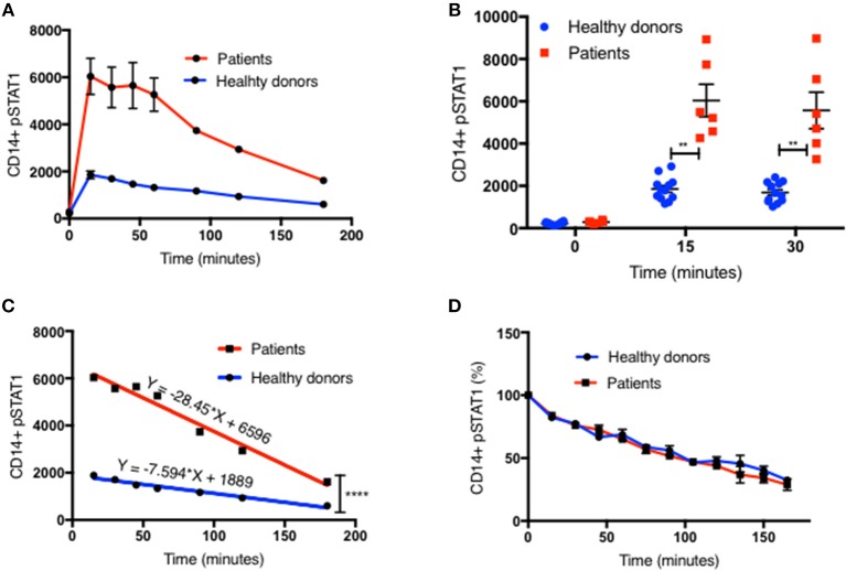
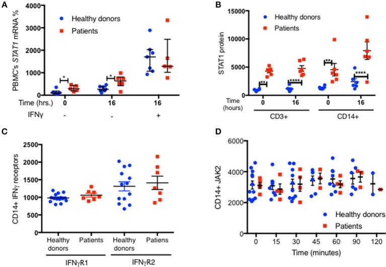
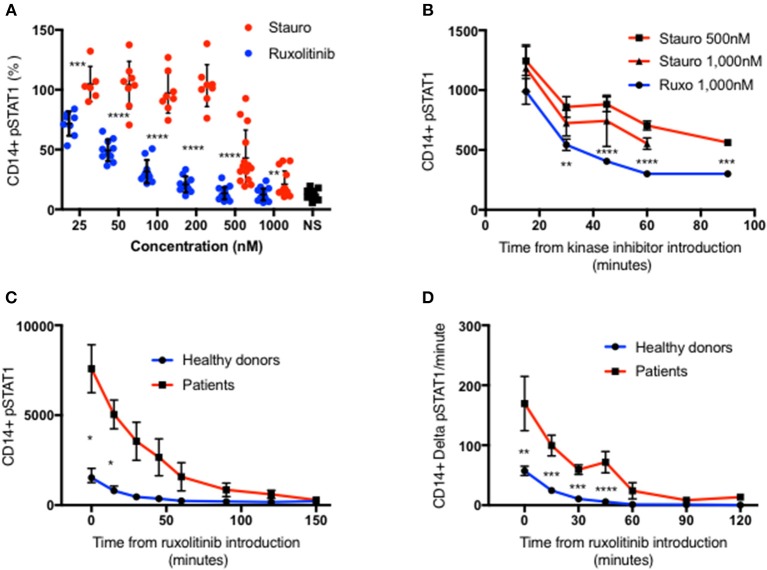
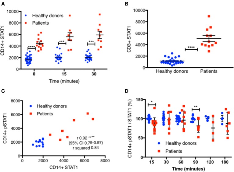
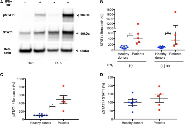
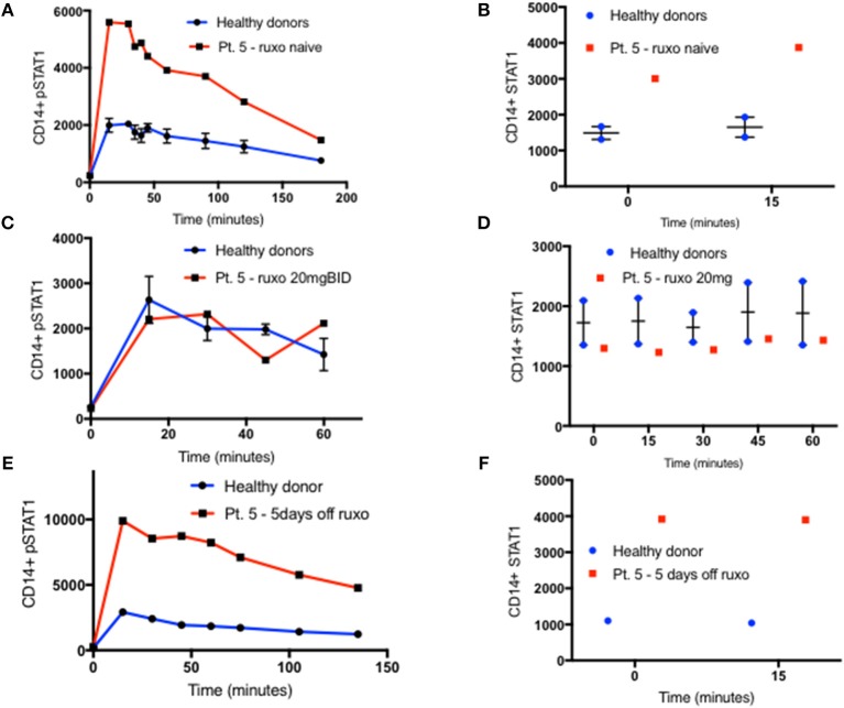
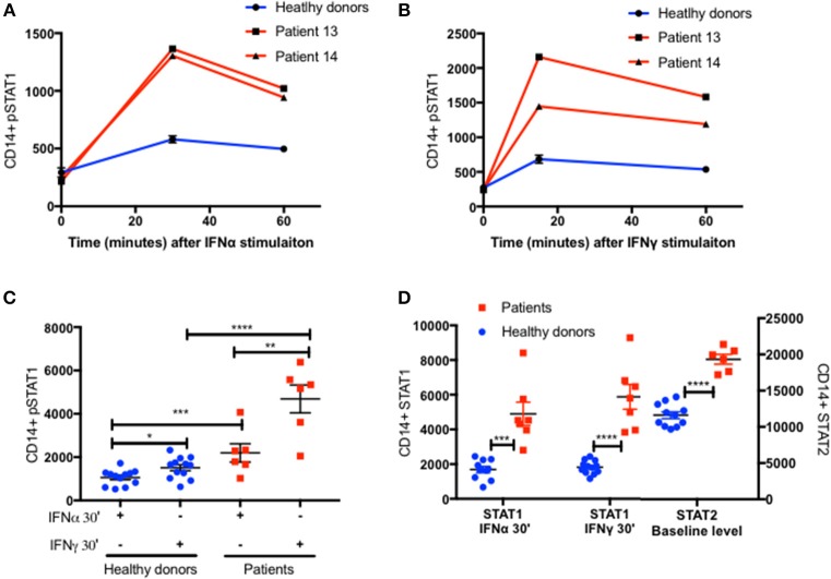
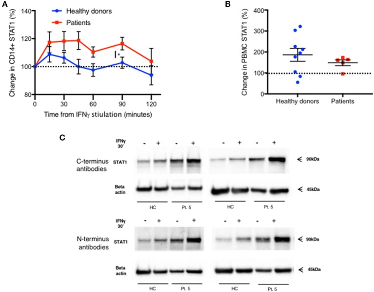
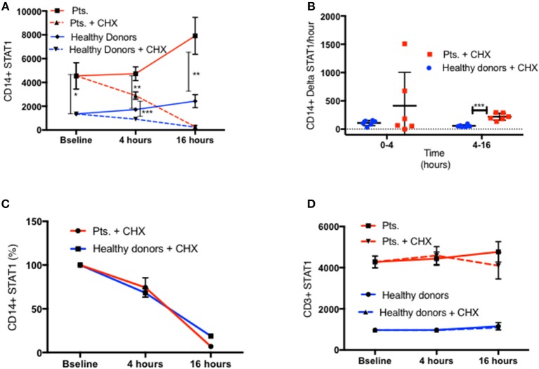
Signal transducer and activator of transcription gain of function (GOF) pathogenic variants have been associated with increased levels of phosphorylated STAT1 and STAT1-dependent cellular responses. Delayed dephosphorylation was proposed as the underlying mechanism leading to the characteristically raised pSTAT1 levels. We examined the levels of STAT1 protein and message as well as rates of STAT1 phosphorylation, dephosphorylation, and degradation associated with GOF pathogenic variants. Fresh peripheral blood mononuclear cells (PBMC) from 14 STAT1 GOF patients carrying 10 different pathogenic variants in the coiled-coil, DNA binding, and SH2 domains and healthy donors were used to study STAT1 levels and phosphorylation (pSTAT1) following IFNγ and IFNα stimulation. STAT1 protein levels were measured by flow cytometry and immunoblot. mRNA levels were measured using quantitative reverse transcription PCR. STAT1 protein degradation was studied using cycloheximide. Patient IFNγ and IFNα induced peak pSTAT1 was higher than in healthy controls. The velocity of pSTAT1 dephosphorylation after treatment of IFNγ stimulated CD14 monocytes with the Janus Kinase (JAK)-inhibitor ruxolitinib was significantly faster in patient cells. STAT1 protein levels in patient CD14 monocytes and CD3 T cells were higher than in healthy donors. There was a strong and positive correlation between CD14 STAT1 protein levels and peak pSTAT1 levels. Patient fresh PBMC mRNA levels were increased at rest and after 16 h of incubation. STAT1 protein degradation was similar in patient and healthy volunteer cells. Patient IFNγ receptors 1 and 2 and JAK2 levels were normal. One patient in our cohort was treated with the oral JAK inhibitor ruxolitinib. Treatment was associated with normalization of both STAT1 protein and peak pSTAT1 levels. After JAK inhibitor treatment was stopped the patient's CD14 monocyte STAT1 protein and peak phosphorylation levels increased proportionally. These findings suggest that patients with GOF mutations have higher levels of total STAT1 protein, leading to high levels of pSTAT1 after stimulation, despite rapid STAT1 dephosphorylation and normal degradation.
信号转导子和转录激活子 3 获得性功能(GOF)致病性变异与磷酸化 STAT1 水平升高和 STAT1 依赖性细胞反应有关。提出磷酸化 STAT1 水平升高的潜在机制是去磷酸化延迟。我们检查了与 GOF 致病性变异相关的 STAT1 蛋白和信使 RNA 水平以及 STAT1 磷酸化、去磷酸化和降解的速率。使用来自 14 名携带卷曲螺旋、DNA 结合和 SH2 结构域中 10 种不同致病性变异的 STAT1 GOF 患者和健康供体的新鲜外周血单核细胞(PBMC),研究 IFNγ 和 IFNα 刺激后 STAT1 水平和磷酸化(pSTAT1)。通过流式细胞术和免疫印迹测量 STAT1 蛋白水平。使用定量逆转录 PCR 测量 mRNA 水平。使用环已酰亚胺研究 STAT1 蛋白降解。患者 IFNγ 和 IFNα 诱导的 pSTAT1 峰值高于健康对照组。用 Janus Kinase(JAK)抑制剂 ruxolitinib 处理 IFNγ 刺激的 CD14 单核细胞后,pSTAT1 去磷酸化的速度在患者细胞中明显更快。患者 CD14 单核细胞和 CD3 T 细胞中的 STAT1 蛋白水平高于健康供体。CD14 STAT1 蛋白水平与 pSTAT1 峰值之间存在强烈的正相关。患者新鲜 PBMC 在休息时和孵育 16 小时后 mRNA 水平增加。STAT1 蛋白降解在患者和健康志愿者细胞中相似。患者 IFNγ 受体 1 和 2 和 JAK2 水平正常。我们队列中的一名患者接受了口服 JAK 抑制剂 ruxolitinib 的治疗。治疗与 STAT1 蛋白和 pSTAT1 峰值水平的正常化相关。停止 JAK 抑制剂治疗后,患者的 CD14 单核细胞 STAT1 蛋白和峰磷酸化水平成比例增加。这些发现表明,GOF 突变患者具有更高水平的总 STAT1 蛋白,导致刺激后 pSTAT1 水平升高,尽管 STAT1 去磷酸化迅速且正常降解。