Janelia Research Campus, Howard Hughes Medical Institute, Ashburn, VA, USA.
Department of Neurobiology, Stanford University, Stanford, CA, USA.
Nat Methods. 2019 Aug;16(8):778-786. doi: 10.1038/s41592-019-0493-9. Epub 2019 Jul 29.
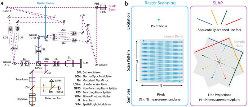
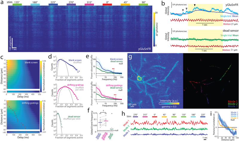
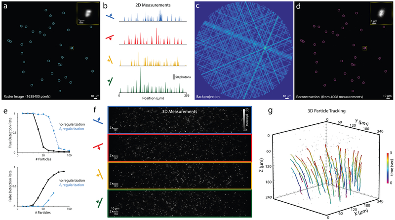
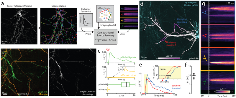
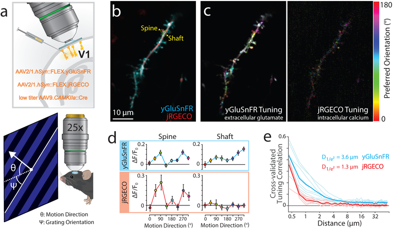
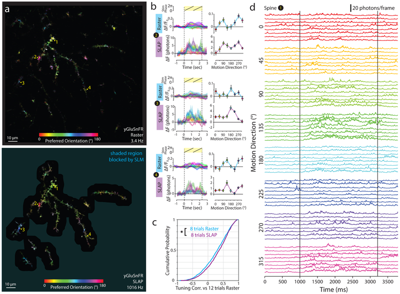
Point-scanning two-photon microscopy enables high-resolution imaging within scattering specimens such as the mammalian brain, but sequential acquisition of voxels fundamentally limits its speed. We developed a two-photon imaging technique that scans lines of excitation across a focal plane at multiple angles and computationally recovers high-resolution images, attaining voxel rates of over 1 billion Hz in structured samples. Using a static image as a prior for recording neural activity, we imaged visually evoked and spontaneous glutamate release across hundreds of dendritic spines in mice at depths over 250 µm and frame rates over 1 kHz. Dendritic glutamate transients in anesthetized mice are synchronized within spatially contiguous domains spanning tens of micrometers at frequencies ranging from 1-100 Hz. We demonstrate millisecond-resolved recordings of acetylcholine and voltage indicators, three-dimensional single-particle tracking and imaging in densely labeled cortex. Our method surpasses limits on the speed of raster-scanned imaging imposed by fluorescence lifetime.
点扫描双光子显微镜能够在散射样本(如哺乳动物大脑)中实现高分辨率成像,但体素的顺序采集从根本上限制了其速度。我们开发了一种双光子成像技术,该技术可以在多个角度扫描焦平面上的激发线,并通过计算恢复高分辨率图像,在结构样本中实现超过 10 亿 Hz 的体素率。我们使用静态图像作为记录神经活动的先验,在超过 250 µm 的深度和超过 1 kHz 的帧率下,对小鼠数百个树突棘中的视觉诱发和自发谷氨酸释放进行成像。麻醉小鼠的树突谷氨酸瞬变在数十微米的空间连续区域内以 1-100 Hz 的频率同步。我们演示了毫秒分辨率的乙酰胆碱和电压指示剂记录、在密集标记的皮质中的三维单颗粒跟踪和成像。我们的方法超越了荧光寿命对光栅扫描成像速度的限制。