De Oliveira Ernane Lacerda, De Carvalho Paulo Sergio Perri, Da Silva Thiago Bezerra
Department of Implantology, São Leopoldo Mandic, São Paulo, Brazil.
Department of Implantology, CPO São Leopoldo Mandic, Campinas, São Paulo, Brazil.
J Indian Soc Periodontol. 2019 Jul-Aug;23(4):351-355. doi: 10.4103/jisp.jisp_111_18.
The last few years have detailed a number of surgical materials and techniques to stimulate guided bone regeneration (GBR). Polypropylene has been used as a mechanical barrier, intentionally designed to be exposed to the oral environment, isolating the regeneration area, and allowing the blood clot to remain protected in a confined space while pluripotent mesenchymal cells regenerate the alveolar bone tissue.
Due to the lack of studies on polypropylene barriers (PB) (Bone Heal- Bone heal ind. e Com. LTDA - São Paulo, Brazil), this study aimed to evaluate the histological repair process of critical defects (7 mm) made in the rodent cranial vaults comparing its efficacy in GBR and modified GBR.
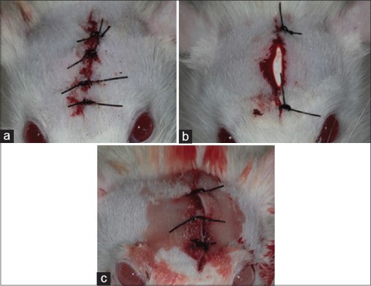
A total of 30 rats were divided into three groups. The control group consisted of 10 rats, wounds covered with just a blood clot. The second group consisted of an exposed/uncoated PB at the edges of the wound, and it was removed after 3 days. The third group used submerged PB with coaptation of the wound edges, and it was not removed. Five animals of each group were euthanized at 30 and 90 days postoperative and submitted to microscopic analysis and histomorphometric evaluation.
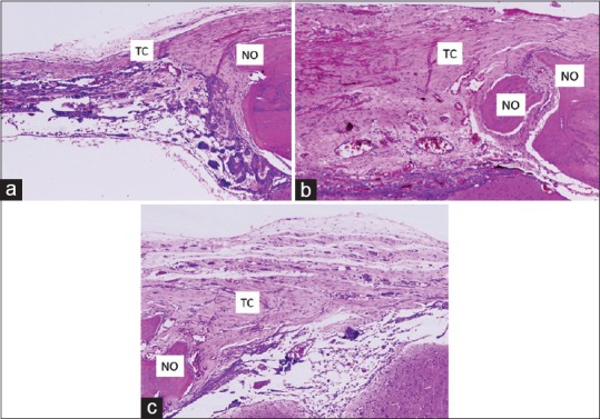
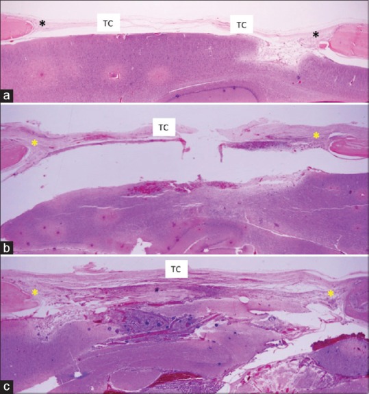
Modified GBR (membrane exposed to the oral medium) provided earlier tissue organization at 30 days; however, the third group presented better bone neoformation at 90 days.
Modified GBR provided earlier tissue organization compared with the control group, as well as promoting improved bone neoformation, while regeneration with the submerged membrane presented better bone neoformation in the long term.
过去几年详细介绍了多种用于促进引导性骨再生(GBR)的手术材料和技术。聚丙烯已被用作机械屏障,特意设计用于暴露于口腔环境中,隔离再生区域,并在多能间充质细胞再生牙槽骨组织时,使血凝块在封闭空间内得到保护。
由于缺乏关于聚丙烯屏障(PB)(巴西圣保罗的Bone Heal - Bone heal ind. e Com. LTDA公司生产)的研究,本研究旨在评估在啮齿动物颅顶制造的临界缺损(7毫米)的组织学修复过程,比较其在引导性骨再生和改良引导性骨再生中的疗效。
总共30只大鼠被分为三组。对照组由10只大鼠组成,伤口仅覆盖血凝块。第二组在伤口边缘使用暴露/未涂层的PB,3天后移除。第三组使用与伤口边缘贴合的埋入式PB,且未移除。每组五只动物在术后30天和90天实施安乐死,并进行显微镜分析和组织形态计量学评估。
改良引导性骨再生(膜暴露于口腔介质)在30天时能更早实现组织构建;然而,第三组在90天时骨新生情况更佳。
与对照组相比,改良引导性骨再生能更早实现组织构建,同时促进更好的骨新生,而埋入式膜再生从长期来看骨新生情况更佳。