Wittfoth Christin, Harzsch Steffen, Wolff Carsten, Sombke Andy
1Department of Cytology and Evolutionary Biology, Zoological Institute and Museum, University of Greifswald, Soldmannstr. 23, 17487 Greifswald, Germany.
2Department of Biology, Comparative Zoology, Humboldt University Berlin, Philippstr. 13, 10115 Berlin, Germany.
Front Zool. 2019 Jul 26;16:30. doi: 10.1186/s12983-019-0330-0. eCollection 2019.
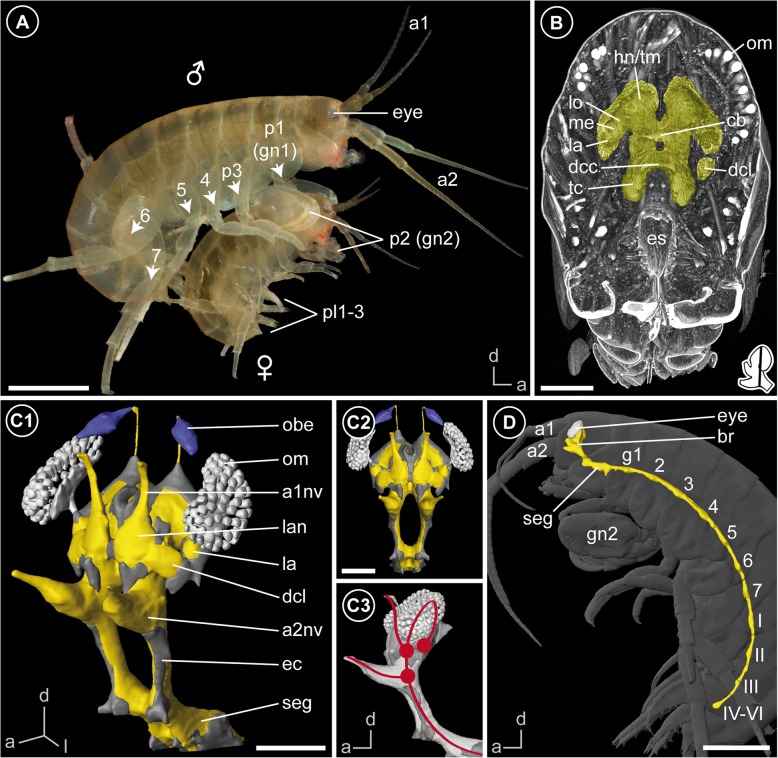
Over the last years, the amphipod crustacean has developed into an attractive marine animal model for evolutionary developmental studies that offers several advantages over existing experimental organisms. It is easy to rear in laboratory conditions with embryos available year-round and amenable to numerous kinds of embryological and functional genetic manipulations. However, beyond these developmental and genetic analyses, research on the architecture of its nervous system is fragmentary. In order to provide a first neuroanatomical atlas of the brain, we investigated using immunohistochemical labelings combined with laser-scanning microscopy, X-ray microcomputed tomography, histological sectioning and 3D reconstructions.
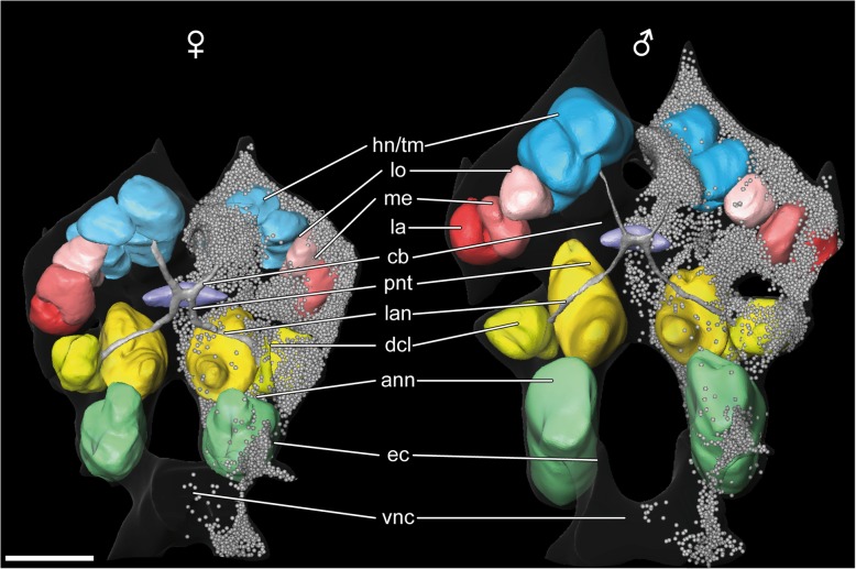
As in most amphipod crustaceans, the brain is dorsally bent out of the body axis with downward oriented lateral hemispheres of the protocerebrum. It comprises almost all prominent neuropils that are part of the suggested ground pattern of malacostracan crustaceans (except the lobula plate and projection neuron tract neuropil). Beyond a general uniformity of these neuropils, the brain of is characterized by an elaborated central complex and a modified lamina (first order visual neuropil), which displays a chambered appearance. In the light of a recent analysis on photoreceptor projections in , the observed architecture of the lamina corresponds to specialized photoreceptor terminals. Furthermore, in contrast to previous descriptions of amphipod brains, we suggest the presence of a poorly differentiated hemiellipsoid body and an inner chiasm and critically discuss these aspects.
Despite a general uniformity of amphipod brains, there is also a certain degree of variability in architecture and size of different neuropils, reflecting various ecologies and life styles of different species. In contrast to other amphipods, the brain of does not display any striking modifications or bias towards processing one particular sensory modality. Thus, we conclude that this brain represents a common type of an amphipod brain. Considering various established protocols for analyzing and manipulating , this organism is a suitable model to gain deeper understanding of brain anatomy e.g. by using connectome approaches, and this study can serve as first solid basis for following studies.
在过去几年中,这种双足甲壳类动物已发展成为一种有吸引力的海洋动物模型,用于进化发育研究,与现有的实验生物相比具有诸多优势。它易于在实验室条件下饲养,全年都有胚胎,并且适合进行多种胚胎学和功能遗传学操作。然而,除了这些发育和遗传分析之外,对其神经系统结构的研究还很零散。为了提供第一份脑的神经解剖图谱,我们结合激光扫描显微镜、X射线微计算机断层扫描、组织切片和三维重建,使用免疫组织化学标记进行了研究。
与大多数双足甲壳类动物一样,脑背向弯曲离开身体轴线,原脑的侧半球向下。它包含几乎所有突出的神经纤维网,这些神经纤维网是软甲亚纲甲壳动物假定基本模式的一部分(除小叶板和投射神经元束神经纤维网外)。除了这些神经纤维网的一般一致性之外,该动物的脑的特征在于一个精细的中央复合体和一个经过修饰的层板(一级视觉神经纤维网),其呈现出腔室状外观。根据最近对该动物光感受器投射的分析,观察到的层板结构对应于特化的光感受器终端。此外,与之前对双足类动物脑的描述相反,我们认为存在一个分化程度较低的半椭球体和一个内交叉,并对这些方面进行了批判性讨论。
尽管双足类动物的脑具有一般的一致性,但不同神经纤维网的结构和大小也存在一定程度的变异性,反映了不同物种的各种生态和生活方式。与其他双足类动物不同,该动物的脑没有表现出任何显著的改变或对处理一种特定感觉模态的偏向。因此,我们得出结论,这种脑代表了双足类动物脑的一种常见类型。考虑到用于分析和操作该动物的各种既定方案,这种生物是深入了解脑解剖学的合适模型,例如通过使用连接体方法,并且这项研究可以作为后续研究的第一个坚实基础。