Monash Alfred Psychiatry Research Centre, Monash University Central Clinical School, Melbourne, Victoria, Australia.
Epworth Centre for Innovation in Mental Health, Epworth Healthcare, The Epworth Clinic, Camberwell, Victoria, Australia.
PLoS One. 2019 Aug 6;14(8):e0203096. doi: 10.1371/journal.pone.0203096. eCollection 2019.
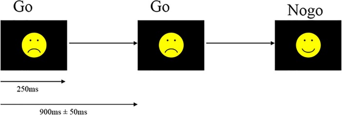
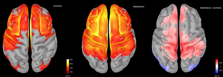
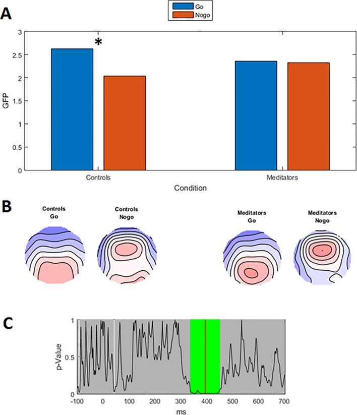
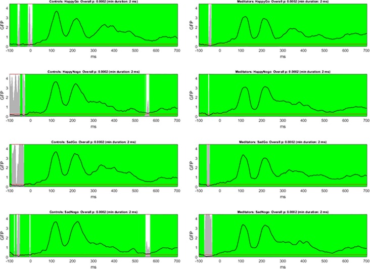
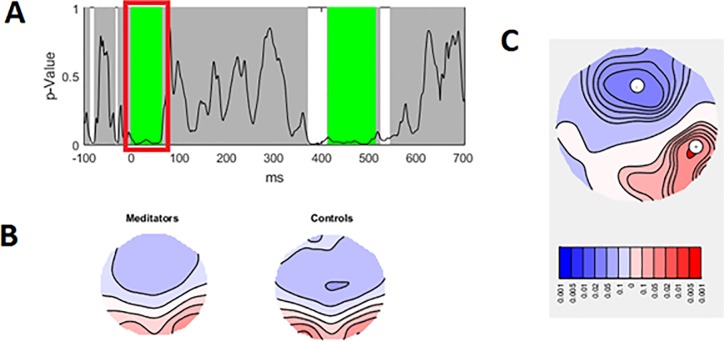
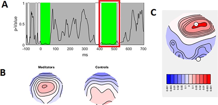
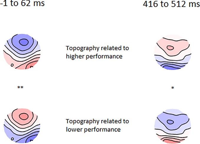
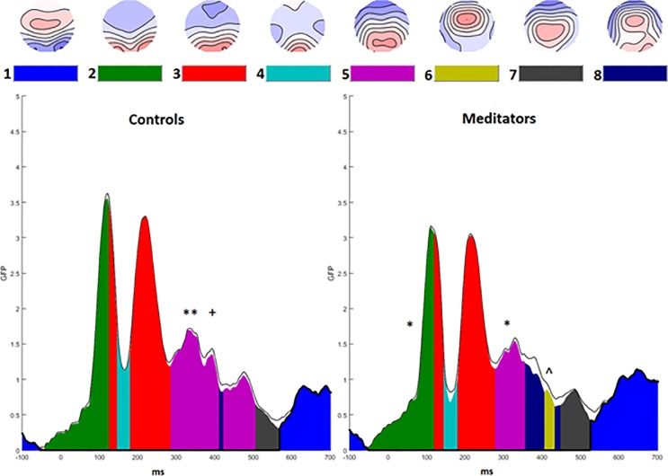
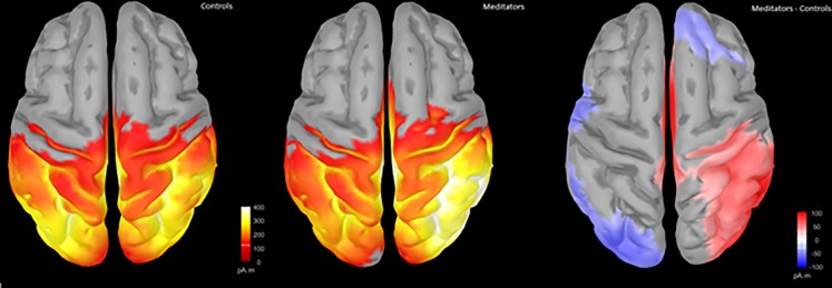
Attention is vital for optimal behavioural performance in every-day life. Mindfulness meditation has been shown to enhance attention. However, the components of attention altered by meditation and the related neural activities are underexplored. In particular, the contributions of inhibitory processes and sustained attention are not well understood. To address these points, 34 meditators were compared to 28 age and gender matched controls during electroencephalography (EEG) recordings of neural activity during a Go/Nogo response inhibition task. This task generates a P3 event related potential, which is related to response inhibition processes in Nogo trials, and attention processes across both trial types. Compared with controls, meditators were more accurate at responding to Go and Nogo trials. Meditators showed a more frontally distributed P3 to both Go and Nogo trials, suggesting more frontal involvement in sustained attention rather than activity specific to response inhibition. Unexpectedly, meditators also showed increased positivity over the right parietal cortex prior to visual information reaching the occipital cortex (during the pre-C1 window). Both results were positively related to increased accuracy across both groups. The results suggest that meditators show altered engagement of neural regions related to attention, including both higher order processes generated by frontal regions, and sensory anticipation processes generated by poster regions. This activity may reflect an increased capacity to modulate a range of neural processes in order to meet task requirements. This increased capacity may underlie the improved attentional function observed in mindfulness meditators.
注意力对于日常生活中的最佳行为表现至关重要。正念冥想已被证明可以提高注意力。然而,冥想改变的注意力成分和相关的神经活动仍未得到充分探索。特别是,抑制过程和持续注意力的贡献还不太清楚。为了解决这些问题,我们在脑电图(EEG)记录中比较了 34 名冥想者和 28 名年龄和性别匹配的对照组在执行 Go/Nogo 反应抑制任务时的神经活动。该任务产生了 P3 事件相关电位,与 Nogo 试验中的反应抑制过程以及两种试验类型的注意力过程有关。与对照组相比,冥想者在对 Go 和 Nogo 试验的反应更加准确。冥想者在 Go 和 Nogo 试验中均显示出更分布于额叶的 P3,这表明在持续注意力方面涉及更多的额叶,而不是特定于反应抑制的活动。出乎意料的是,冥想者在视觉信息到达枕叶皮质之前(在 C1 前窗期间),右顶叶皮质上也显示出增加的正性。这两个结果均与两组的准确性提高呈正相关。结果表明,冥想者表现出与注意力相关的神经区域的改变参与,包括额叶区域产生的更高阶过程,以及后叶区域产生的感觉预期过程。这种活动可能反映了增强调节一系列神经过程以满足任务要求的能力。这种增强的能力可能是正念冥想者观察到注意力功能改善的基础。