• 文献检索
  • 文档翻译
  • 深度研究
  • 学术资讯
  • Suppr Zotero 插件Zotero 插件
  • 邀请有礼
  • 套餐&价格
  • 历史记录
应用&插件
Suppr Zotero 插件Zotero 插件浏览器插件Mac 客户端Windows 客户端微信小程序
定价
高级版会员购买积分包购买API积分包
服务
文献检索文档翻译深度研究API 文档MCP 服务
关于我们
关于 Suppr公司介绍联系我们用户协议隐私条款
关注我们

Suppr 超能文献

核心技术专利:CN118964589B侵权必究
粤ICP备2023148730 号-1Suppr @ 2026

文献检索

告别复杂PubMed语法,用中文像聊天一样搜索,搜遍4000万医学文献。AI智能推荐,让科研检索更轻松。

立即免费搜索

文件翻译

保留排版,准确专业,支持PDF/Word/PPT等文件格式,支持 12+语言互译。

免费翻译文档

深度研究

AI帮你快速写综述,25分钟生成高质量综述,智能提取关键信息,辅助科研写作。

立即免费体验

开角型青光眼视网膜神经纤维层缺损的位置及相关因素

Location of Retinal Nerve Fiber Layer Defects in Open-angle Glaucoma and Associated Factors.

作者信息

Kim Han Ul, Na Kyeong Ik

机构信息

Department of Ophthalmology, Kangdong Sacred Heart Hospital, Hallym University College of Medicine, Seoul, Korea.

出版信息

Korean J Ophthalmol. 2019 Aug;33(4):379-385. doi: 10.3341/kjo.2018.0094.

DOI:10.3341/kjo.2018.0094
PMID:31389215
原文链接:https://pmc.ncbi.nlm.nih.gov/articles/PMC6685826/
Abstract

PURPOSE

To investigate the location of retinal nerve fiber layer defects (RNFLDs) in open-angle glaucoma and the differences in systemic and ocular factors between superotemporal and inferotemporal RNFLDs.

METHODS

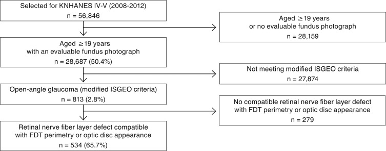
We performed a retrospective review of the 2008 to 2012 data from the Korea National Health and Nutrition Examination Survey. Subjects aged ≥19 years with an evaluable fundus photograph of at least one eye were enrolled, and open-angle glaucoma was diagnosed according to modified International Society of Geographical and Epidemiological Ophthalmology criteria. In subjects with open-angle glaucoma, locations of RNFLDs were evaluated, and systemic and ocular factors were compared between the bilateral superotemporal RNFLD group and bilateral inferotemporal RNFLD group.

RESULTS

A total of 534 subjects had open-angle glaucoma with RNFLDs. The unilateral inferotemporal region (25.8%) was the most common location for RNFLDs, followed by the unilateral superotemporal region (24.4%). Multivariate analysis revealed that hypertension was more significantly associated ( = 0.048) with the bilateral superotemporal RNFLD group than with the bilateral inferotemporal RNFLD group.

CONCLUSIONS

Superotemporal RNFLDs are more related to hypertension than are inferotemporal RNFLDs.

摘要

目的

研究开角型青光眼视网膜神经纤维层缺损(RNFLDs)的位置,以及颞上和颞下RNFLDs在全身和眼部因素方面的差异。

方法

我们对韩国国家健康与营养检查调查2008年至2012年的数据进行了回顾性分析。纳入年龄≥19岁且至少有一只眼睛有可评估眼底照片的受试者,并根据改良的国际地理和流行病学眼科学会标准诊断开角型青光眼。在开角型青光眼患者中,评估RNFLDs的位置,并比较双侧颞上RNFLD组和双侧颞下RNFLD组的全身和眼部因素。

结果

共有534例开角型青光眼患者伴有RNFLDs。单侧颞下区域(25.8%)是RNFLDs最常见的位置,其次是单侧颞上区域(24.4%)。多因素分析显示,与双侧颞下RNFLD组相比,高血压与双侧颞上RNFLD组的相关性更强(P = 0.048)。

结论

与颞下RNFLDs相比,颞上RNFLDs与高血压的关系更为密切。

https://cdn.ncbi.nlm.nih.gov/pmc/blobs/51da/6685826/a6c3325de3bb/kjo-33-379-g001.jpg

相似文献

1
Location of Retinal Nerve Fiber Layer Defects in Open-angle Glaucoma and Associated Factors.开角型青光眼视网膜神经纤维层缺损的位置及相关因素
Korean J Ophthalmol. 2019 Aug;33(4):379-385. doi: 10.3341/kjo.2018.0094.
2
Agreement between spectral domain optical coherence tomography and retinal nerve fiber layer photography in chinese.中文的谱域光相干断层扫描和视网膜神经纤维层照相的一致性。
J Glaucoma. 2012 Apr-May;21(4):228-33. doi: 10.1097/IJG.0b013e3182070cc6.
3
Evaluation of dynamic thiol-disulfide homeostasis in glaucoma patients and the correlation with retinal nerve fiber layer analysis.青光眼患者动态硫醇-二硫键稳态评估及其与视网膜神经纤维层分析的相关性
Eur J Ophthalmol. 2020 Jul;30(4):690-699. doi: 10.1177/1120672119839582. Epub 2019 Apr 12.
4
Circumpapillary microperimetry to detect glaucoma: a pilot study for sector-based comparison to circumpapillary retinal nerve fiber layer measurement.检测青光眼的视乳头周围微视野检查:一项基于象限与视乳头周围视网膜神经纤维层测量进行比较的初步研究。
Int Ophthalmol. 2019 Jan;39(1):127-136. doi: 10.1007/s10792-017-0796-8. Epub 2017 Dec 16.
5
Detection of localized retinal nerve fiber layer defects with posterior pole asymmetry analysis of spectral domain optical coherence tomography.利用频域光学相干断层扫描的后极点不对称分析检测局部视网膜神经纤维层缺损。
Invest Ophthalmol Vis Sci. 2012 Jul 1;53(8):4347-53. doi: 10.1167/iovs.12-9673.
6
Measurement of peripapillary retinal nerve fiber layer volume in glaucoma.青光眼患者视乳头周围视网膜神经纤维层容积的测量
Am J Ophthalmol. 2000 May;129(5):599-607. doi: 10.1016/s0002-9394(99)00464-x.
7
Ocular characteristics associated with the location of focal lamina cribrosa defects in open-angle glaucoma patients.与开角型青光眼患者视盘筛板局限性缺损位置相关的眼部特征。
Eye (Lond). 2017 Apr;31(4):578-587. doi: 10.1038/eye.2016.270. Epub 2016 Dec 9.
8
[Correlation between the retinal nerve fiber layer defects (RNFLD) and visual field defects in primary open-angle glaucoma].[原发性开角型青光眼视网膜神经纤维层缺损(RNFLD)与视野缺损的相关性]
Zhonghua Yan Ke Za Zhi. 1990 Jul;26(4):205-8.
9
Progressive Decrease of Peripapillary Angioflow Vessel Density During Structural and Visual Field Progression in Early Primary Open-angle Glaucoma.早期原发性开角型青光眼结构和视野进展过程中视乳头周围血管血流密度的逐渐降低
J Glaucoma. 2017 Jul;26(7):661-664. doi: 10.1097/IJG.0000000000000695.
10
Location of Disc Hemorrhage and Direction of Progression in Glaucomatous Retinal Nerve Fiber Layer Defects.盘膜下出血位置与青光眼性视网膜神经纤维层缺损进展方向。
J Glaucoma. 2018 Jun;27(6):504-510. doi: 10.1097/IJG.0000000000000945.

引用本文的文献

1
Vascular Density and Retinal Structure Relationships Across Sectors and Regions of the Optic Nerve in Patients With Primary Open Angle Glaucoma.原发性开角型青光眼患者视神经各扇区和区域的血管密度与视网膜结构关系
J Glaucoma. 2025 Apr 1;34(4):267-274. doi: 10.1097/IJG.0000000000002536. Epub 2025 Jan 16.
2
Retinal nerve fiber layer defects and chronic kidney disease: the Kailuan Eye Study.视网膜神经纤维层缺损与慢性肾脏病:开滦眼病研究
Int J Ophthalmol. 2024 Sep 18;17(9):1696-1706. doi: 10.18240/ijo.2024.09.18. eCollection 2024.
3
Corneal endothelium, retinal nerve fiber layer, ganglion cell complex, and perimetry measurements in normal eyes and those with primary open-angle glaucoma.

本文引用的文献

1
Prevalence of retinal nerve fiber layer defects: The Korea National Health and Nutrition Examination Survey 2008-2012.视网膜神经纤维层缺损的患病率:2008 - 2012年韩国国家健康与营养检查调查
PLoS One. 2017 Oct 5;12(10):e0186032. doi: 10.1371/journal.pone.0186032. eCollection 2017.
2
Prevalence, Awareness, and Risk Factors of Primary Open-Angle Glaucoma: Korea National Health and Nutrition Examination Survey 2008-2011.原发性开角型青光眼的流行率、知晓率和危险因素:韩国 2008-2011 年国家健康和营养调查。
Ophthalmology. 2016 Mar;123(3):532-41. doi: 10.1016/j.ophtha.2015.11.004. Epub 2015 Dec 30.
3
Retinal nerve fiber layer loss is associated with urinary albumin excretion in patients with type 2 diabetes.
正常眼与原发性开角型青光眼患者的角膜内皮、视网膜神经纤维层、神经节细胞复合体及视野测量
Med Hypothesis Discov Innov Ophthalmol. 2022 Sep 23;11(2):85-91. doi: 10.51329/mehdiophthal1450. eCollection 2022 Summer.
视网膜神经纤维层丢失与 2 型糖尿病患者的尿白蛋白排泄量有关。
Ophthalmology. 2015 May;122(5):976-81. doi: 10.1016/j.ophtha.2015.01.001. Epub 2015 Feb 7.
4
Diabetes mellitus as a risk factor for open-angle glaucoma: a systematic review and meta-analysis.糖尿病作为开角型青光眼的一个危险因素:一项系统评价和荟萃分析。
PLoS One. 2014 Aug 19;9(8):e102972. doi: 10.1371/journal.pone.0102972. eCollection 2014.
5
Predictors for incidence of primary open-angle glaucoma in a South Indian population: the Chennai eye disease incidence study.在一个印度南部人群中预测原发性开角型青光眼发病的因素:Chennai 眼病发病研究。
Ophthalmology. 2014 Jul;121(7):1370-6. doi: 10.1016/j.ophtha.2014.01.014. Epub 2014 Mar 18.
6
Glaucomatous damage of the macula.黄斑青光眼损害。
Prog Retin Eye Res. 2013 Jan;32:1-21. doi: 10.1016/j.preteyeres.2012.08.003. Epub 2012 Sep 17.
7
Comparison of the progression rates of the superior, inferior, and both hemifield defects in normal-tension glaucoma patients.正常眼压性青光眼患者的上方、下方和双侧视野缺损进展率比较。
Am J Ophthalmol. 2012 Dec;154(6):958-968.e1. doi: 10.1016/j.ajo.2012.05.025. Epub 2012 Sep 7.
8
Intraocular pressure correlated with arterial blood pressure: the beijing eye study.眼压与动脉血压的相关性:北京眼病研究
Am J Ophthalmol. 2007 Sep;144(3):461-2. doi: 10.1016/j.ajo.2007.05.013.
9
The eye in hypertension.高血压患者的眼睛
Lancet. 2007 Feb 3;369(9559):425-35. doi: 10.1016/S0140-6736(07)60198-6.
10
Risk factors for open-angle glaucoma in a Japanese population: the Tajimi Study.日本人群开角型青光眼的危险因素:多治见研究
Ophthalmology. 2006 Sep;113(9):1613-7. doi: 10.1016/j.ophtha.2006.03.059. Epub 2006 Jul 7.