• 文献检索
  • 文档翻译
  • 深度研究
  • 学术资讯
  • Suppr Zotero 插件Zotero 插件
  • 邀请有礼
  • 套餐&价格
  • 历史记录
应用&插件
Suppr Zotero 插件Zotero 插件浏览器插件Mac 客户端Windows 客户端微信小程序
定价
高级版会员购买积分包购买API积分包
服务
文献检索文档翻译深度研究API 文档MCP 服务
关于我们
关于 Suppr公司介绍联系我们用户协议隐私条款
关注我们

Suppr 超能文献

核心技术专利:CN118964589B侵权必究
粤ICP备2023148730 号-1Suppr @ 2026

文献检索

告别复杂PubMed语法,用中文像聊天一样搜索,搜遍4000万医学文献。AI智能推荐,让科研检索更轻松。

立即免费搜索

文件翻译

保留排版,准确专业,支持PDF/Word/PPT等文件格式,支持 12+语言互译。

免费翻译文档

深度研究

AI帮你快速写综述,25分钟生成高质量综述,智能提取关键信息,辅助科研写作。

立即免费体验

应用戈勃域光学相干显微镜对人供体角膜内皮的定量评估。

Quantitative assessment of human donor corneal endothelium with Gabor domain optical coherence microscopy.

机构信息

The Institute of Optics, Univ. of Rochester, United States.

Univ. of Rochester, United States.

出版信息

J Biomed Opt. 2019 Aug;24(8):1-9. doi: 10.1117/1.JBO.24.8.085001.

DOI:10.1117/1.JBO.24.8.085001
PMID:31389221
原文链接:https://pmc.ncbi.nlm.nih.gov/articles/PMC6682832/
Abstract

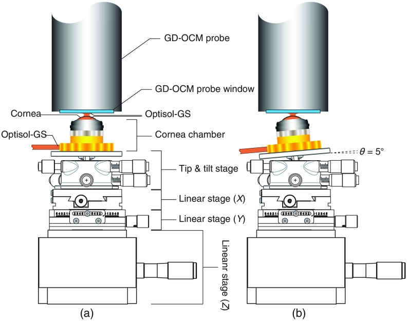
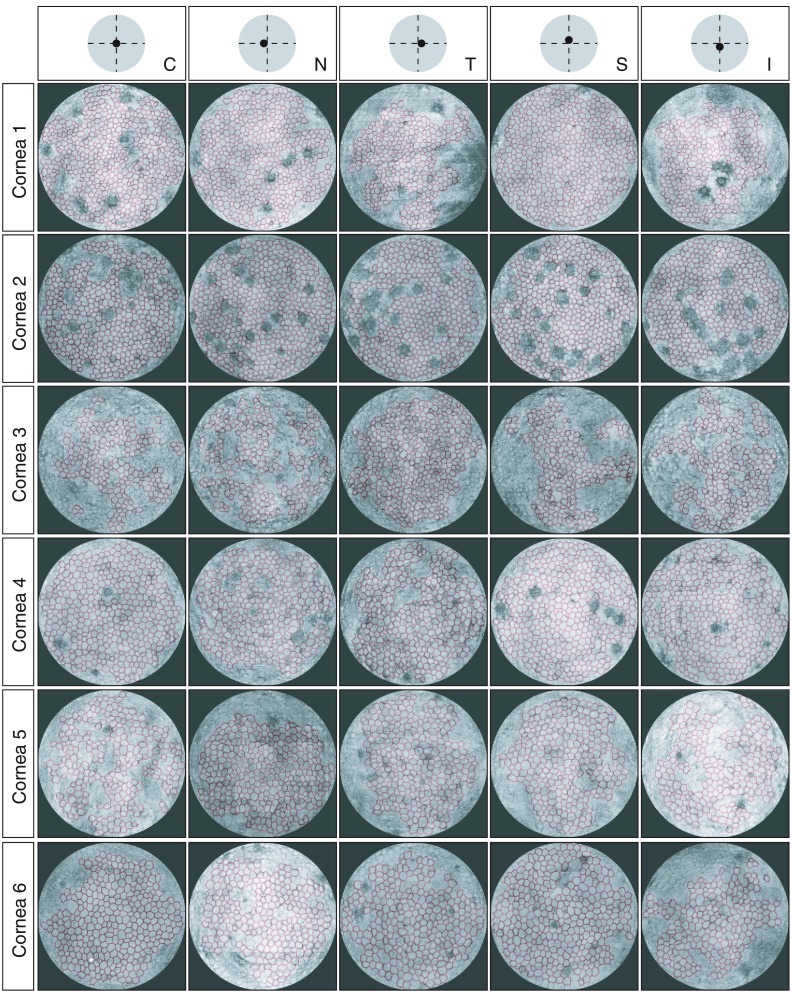
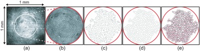
We report on a pathway for Gabor domain optical coherence microscopy (GD-OCM)-based metrology to assess the donor’s corneal endothelial layers ex vivo. Six corneas from the Lions Eye Bank at Albany and Rochester were imaged with GD-OCM. The raw 3-D images of the curved corneas were flattened using custom software to enhance the 2-D visualization of endothelial cells (ECs); then the ECs within a circle of 500-μm-diameter were analyzed using a custom corner method and a cell counting plugin in ImageJ. The EC number, EC area, endothelial cell density (ECD), and polymegethism (CV) were quantified in five different locations for each cornea. The robustness of the method (defined as the repeatability of measurement together with interoperator variability) was evaluated by independently repeating the entire ECD measurement procedure six times by three different examiners. The results from the six corneas show that the current modality reproduces the ECDs with a standard deviation of 2.3% of the mean ECD in every location, whereas the mean ECD across five locations varies by 5.1%. The resolution and imaging area provided through the use of GD-OCM may help to ultimately better assess the quality of donor corneas in transplantation.

摘要

我们报告了一种基于 Gabor 域光学相干显微镜 (GD-OCM) 的计量学方法,用于评估供体角膜内皮层的离体情况。使用 GD-OCM 对来自奥尔巴尼和罗切斯特狮子眼库的六只角膜进行成像。使用定制软件对弯曲角膜的原始 3D 图像进行展平,以增强内皮细胞 (EC) 的 2D 可视化;然后使用定制角方法和 ImageJ 中的细胞计数插件分析直径为 500μm 的圆圈内的 EC。在每个角膜的五个不同位置定量计算 EC 数量、EC 面积、内皮细胞密度 (ECD) 和多形性 (CV)。通过三位不同的检查者独立重复整个 ECD 测量过程六次,评估了该方法的稳健性(定义为测量的可重复性以及操作者之间的变异性)。六个角膜的结果表明,目前的方法在每个位置都能以平均 ECD 的 2.3%的标准差重现 ECD,而五个位置的平均 ECD 则相差 5.1%。通过使用 GD-OCM 提供的分辨率和成像面积,可能有助于最终更好地评估移植供体角膜的质量。

https://cdn.ncbi.nlm.nih.gov/pmc/blobs/76a0/6682832/29c58785c74f/JBO-024-085001-g006.jpg
https://cdn.ncbi.nlm.nih.gov/pmc/blobs/76a0/6682832/183b2ce3a65d/JBO-024-085001-g001.jpg
https://cdn.ncbi.nlm.nih.gov/pmc/blobs/76a0/6682832/a8b96f9d9855/JBO-024-085001-g002.jpg
https://cdn.ncbi.nlm.nih.gov/pmc/blobs/76a0/6682832/a2dea11fa78f/JBO-024-085001-g003.jpg
https://cdn.ncbi.nlm.nih.gov/pmc/blobs/76a0/6682832/cbeb86819a1b/JBO-024-085001-g005.jpg
https://cdn.ncbi.nlm.nih.gov/pmc/blobs/76a0/6682832/29c58785c74f/JBO-024-085001-g006.jpg

相似文献

1
Quantitative assessment of human donor corneal endothelium with Gabor domain optical coherence microscopy.应用戈勃域光学相干显微镜对人供体角膜内皮的定量评估。
J Biomed Opt. 2019 Aug;24(8):1-9. doi: 10.1117/1.JBO.24.8.085001.
2
Unbiased corneal tissue analysis using Gabor-domain optical coherence microscopy and machine learning for automatic segmentation of corneal endothelial cells.利用 Gabor 域光学相干显微镜和机器学习进行无偏角膜组织分析,实现角膜内皮细胞的自动分割。
J Biomed Opt. 2020 Aug;25(9):1-17. doi: 10.1117/1.JBO.25.9.092902.
3
Assessing microstructures of the cornea with Gabor-domain optical coherence microscopy: pathway for corneal physiology and diseases.利用加博尔域光学相干显微镜评估角膜微观结构:角膜生理学与疾病研究途径
Opt Lett. 2015 Mar 15;40(6):1113-6. doi: 10.1364/OL.40.001113.
4
Capabilities of Gabor-domain optical coherence microscopy for the assessment of corneal disease.用于评估角膜疾病的 Gabor 域光学相干显微镜的功能。
J Biomed Opt. 2019 Apr;24(4):1-17. doi: 10.1117/1.JBO.24.4.046002.
5
Evaluation of repeatability for the automatic estimation of endothelial cell density in donor corneas.供体角膜内皮细胞密度自动估计的重复性评估。
Br J Ophthalmol. 2007 Sep;91(9):1213-5. doi: 10.1136/bjo.2006.112763. Epub 2007 Apr 12.
6
In situ ultrahigh-resolution optical coherence tomography characterization of eye bank corneal tissue processed for lamellar keratoplasty.用于板层角膜移植术的眼库角膜组织的原位超高分辨率光学相干断层扫描特征分析
Cornea. 2008 Aug;27(7):802-10. doi: 10.1097/ICO.0b013e318169d6b7.
7
The measurement of corneal thickness from center to limbus in vivo in C57BL/6 and BALB/c mice using two-photon imaging.应用双光子成像技术活体测量 C57BL/6 和 BALB/c 小鼠角膜从中心到边缘的厚度。
Exp Eye Res. 2013 Oct;115:255-62. doi: 10.1016/j.exer.2013.07.025. Epub 2013 Aug 3.
8
Parallelized multi-graphics processing unit framework for high-speed Gabor-domain optical coherence microscopy.用于高速伽柏域光学相干显微镜的并行多图形处理单元框架
J Biomed Opt. 2014 Jul;19(7):71410. doi: 10.1117/1.JBO.19.7.071410.
9
Full-field optical coherence tomography of human donor and pathological corneas.人类供体角膜和病理角膜的全视野光学相干断层扫描
Curr Eye Res. 2015 May;40(5):526-34. doi: 10.3109/02713683.2014.935444. Epub 2014 Sep 24.
10
New parameters in assessment of human donor corneal stroma.评估人类供体角膜基质的新参数。
Acta Ophthalmol. 2017 Jun;95(4):e297-e306. doi: 10.1111/aos.13351. Epub 2017 Jan 30.

引用本文的文献

1
Detection of refractive photokeratectomy traces during eye banking: impossible with organ culture but possible with an active storage machine: case report.眼库中检测屈光性光动力角膜切削术痕迹:器官培养不可能,但主动储存机能:病例报告。
Cell Tissue Bank. 2021 Sep;22(3):479-486. doi: 10.1007/s10561-020-09895-4. Epub 2021 Jan 4.
2
Unbiased corneal tissue analysis using Gabor-domain optical coherence microscopy and machine learning for automatic segmentation of corneal endothelial cells.利用 Gabor 域光学相干显微镜和机器学习进行无偏角膜组织分析,实现角膜内皮细胞的自动分割。
J Biomed Opt. 2020 Aug;25(9):1-17. doi: 10.1117/1.JBO.25.9.092902.
3

本文引用的文献

1
250 kHz, 1.5 µm resolution SD-OCT for cellular imaging of the human cornea.用于人眼角膜细胞成像的250千赫兹、分辨率为1.5微米的频域光学相干断层扫描技术
Biomed Opt Express. 2018 Nov 29;9(12):6569-6583. doi: 10.1364/BOE.9.006569. eCollection 2018 Dec 1.
2
Capabilities of Gabor-domain optical coherence microscopy for the assessment of corneal disease.用于评估角膜疾病的 Gabor 域光学相干显微镜的功能。
J Biomed Opt. 2019 Apr;24(4):1-17. doi: 10.1117/1.JBO.24.4.046002.
3
Comparative Image Atlas of Current and New Technologies in Corneal Donor Tissue Evaluation.
imaging of corneal nerves and cellular structures in mice with Gabor-domain optical coherence microscopy.
利用加博尔域光学相干显微镜对小鼠角膜神经和细胞结构进行成像。
Biomed Opt Express. 2020 Jan 10;11(2):711-724. doi: 10.1364/BOE.379809. eCollection 2020 Feb 1.
角膜供体组织评估中现有技术与新技术的比较图像图谱
Cornea. 2018 Jun;37 Suppl 1:S14-S35. doi: 10.1097/ICO.0000000000001657.
4
Anterior segment optical coherence tomography.眼前节光学相干断层扫描。
Prog Retin Eye Res. 2018 Sep;66:132-156. doi: 10.1016/j.preteyeres.2018.04.002. Epub 2018 Apr 7.
5
high resolution human corneal imaging using full-field optical coherence tomography.使用全场光学相干断层扫描技术进行高分辨率人眼角膜成像。
Biomed Opt Express. 2018 Jan 10;9(2):557-568. doi: 10.1364/BOE.9.000557. eCollection 2018 Feb 1.
6
Visualizing Micro-anatomical Structures of the Posterior Cornea with Micro-optical Coherence Tomography.利用微光学相干断层扫描技术对后角膜的微观解剖结构进行可视化。
Sci Rep. 2017 Sep 7;7(1):10752. doi: 10.1038/s41598-017-11380-0.
7
Conical scan pattern for enhanced visualization of the human cornea using polarization-sensitive OCT.使用偏振敏感光学相干断层扫描技术增强人眼角膜可视化的锥形扫描模式。
Biomed Opt Express. 2017 May 8;8(6):2906-2923. doi: 10.1364/BOE.8.002906. eCollection 2017 Jun 1.
8
Sub-micrometer axial resolution OCT for imaging of the cellular structure of healthy and keratoconic human corneas.用于健康和圆锥角膜人眼角膜细胞结构成像的亚微米轴向分辨率光学相干断层扫描技术
Biomed Opt Express. 2017 Jan 12;8(2):800-812. doi: 10.1364/BOE.8.000800. eCollection 2017 Feb 1.
9
Rapid Warming of Donor Corneas Is Safe and Improves Specular Image Quality.供体角膜快速复温是安全的,并可改善内皮细胞镜图像质量。
Cornea. 2017 May;36(5):581-587. doi: 10.1097/ICO.0000000000001166.
10
Evaluation of a Micro-Optical Coherence Tomography for the Corneal Endothelium in an Animal Model.评价一种用于动物模型角膜内皮的微光学相干断层扫描技术。
Sci Rep. 2016 Jul 15;6:29769. doi: 10.1038/srep29769.