Department of Radiology, Beijing Children's Hospital, Capital Medical University, National Center for Children's Health, No.56 Nanlishi Road, Beijing, 100045, China.
Sino-Dutch Biomedical and Information Engineering School, Northeastern University, No. 3-11 Wenhua Road, Shenyang, China.
BMC Med Imaging. 2019 Aug 8;19(1):63. doi: 10.1186/s12880-019-0355-z.
To investigate the value of predictive nomogram in optimizing computed tomography (CT)-based differential diagnosis of primary progressive pulmonary tuberculosis (TB) from community-acquired pneumonia (CAP) in children.
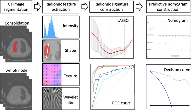
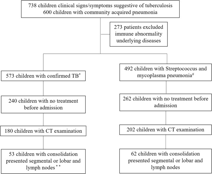
This retrospective study included 53 patients with clinically confirmed pulmonary TB and 62 patients with CAP. Patients were grouped at random according to a 3:1 ratio (primary cohort n = 86, validation cohort n = 29). A total of 970 radiomic features were extracted from CT images and key features were screened out to build radiomic signatures using the least absolute shrinkage and selection operator algorithm. A predictive nomogram was developed based on the signatures and clinical factors, and its performance was assessed by the receiver operating characteristic curve, calibration curve, and decision curve analysis.
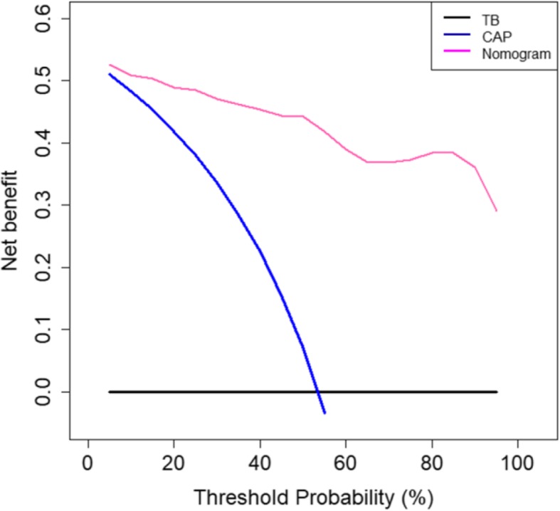
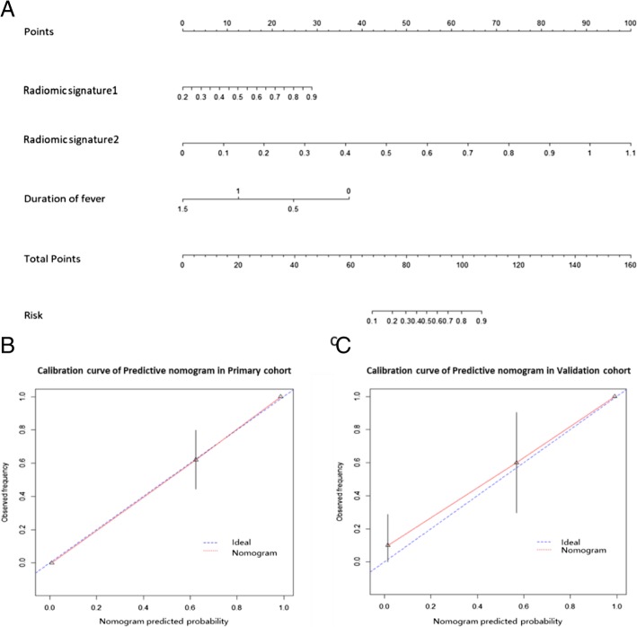
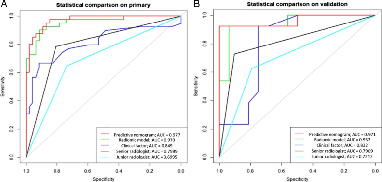
Initially, 5 and 6 key features were selected to establish a radiomic signature from the pulmonary consolidation region (RS1) and a signature from lymph node region (RS2), respectively. A predictive nomogram was built combining RS1, RS2, and a clinical factor (duration of fever). Its classification performance (AUC = 0.971, 95% confidence interval [CI]: 0.912-1) was better than the senior radiologist's clinical judgment (AUC = 0.791, 95% CI: 0.636-0.946), the clinical factor (AUC = 0.832, 95% CI: 0.677-0.987), and the combination of RS1 and RS2 (AUC = 0.957, 95% CI: 0.889-1). The calibration curves indicated a good consistency of the nomogram. Decision curve analysis demonstrated that the nomogram was useful in clinical settings.
A CT-based predictive nomogram was proposed and could be conveniently used to differentiate pulmonary TB from CAP in children.
探讨预测列线图在优化基于计算机断层扫描(CT)的儿童原发性进展性肺结核(TB)与社区获得性肺炎(CAP)鉴别诊断中的价值。
本回顾性研究纳入了 53 例临床确诊肺结核患者和 62 例社区获得性肺炎患者。根据 3:1 的比例(主要队列 n=86,验证队列 n=29)将患者随机分组。从 CT 图像中提取了 970 个放射组学特征,并使用最小绝对值收缩和选择算子算法筛选出关键特征,以构建放射组学特征。根据特征和临床因素建立预测列线图,并通过受试者工作特征曲线、校准曲线和决策曲线分析评估其性能。
最初,从肺实变区(RS1)和淋巴结区(RS2)分别选择 5 个和 6 个关键特征来建立放射组学特征。建立了一个结合 RS1、RS2 和临床因素(发热持续时间)的预测列线图。其分类性能(AUC=0.971,95%置信区间[CI]:0.912-1)优于高级放射科医生的临床判断(AUC=0.791,95%CI:0.636-0.946)、临床因素(AUC=0.832,95%CI:0.677-0.987)和 RS1 和 RS2 的组合(AUC=0.957,95%CI:0.889-1)。校准曲线表明该列线图具有良好的一致性。决策曲线分析表明该列线图在临床环境中具有实用性。
提出了一种基于 CT 的预测列线图,可方便地用于区分儿童肺结核和社区获得性肺炎。