Department of Radiology and Nuclear Medicine, MS Center Amsterdam, Amsterdam Neuroscience, Amsterdam UMC-location VUmc, Amsterdam, the Netherlands.
Department of Radiology and Nuclear Medicine, MS Center Amsterdam, Amsterdam Neuroscience, Amsterdam UMC-location VUmc, Amsterdam, the Netherlands.
Neuroimage Clin. 2019;24:101962. doi: 10.1016/j.nicl.2019.101962. Epub 2019 Aug 6.
Atrophy of the spinal cord is known to occur in multiple sclerosis (MS). The mean upper cervical cord area (MUCCA) can be used to measure this atrophy. Currently, several (semi-)automated methods for MUCCA measurement exist, but validation in clinical magnetic resonance (MR) images is lacking.
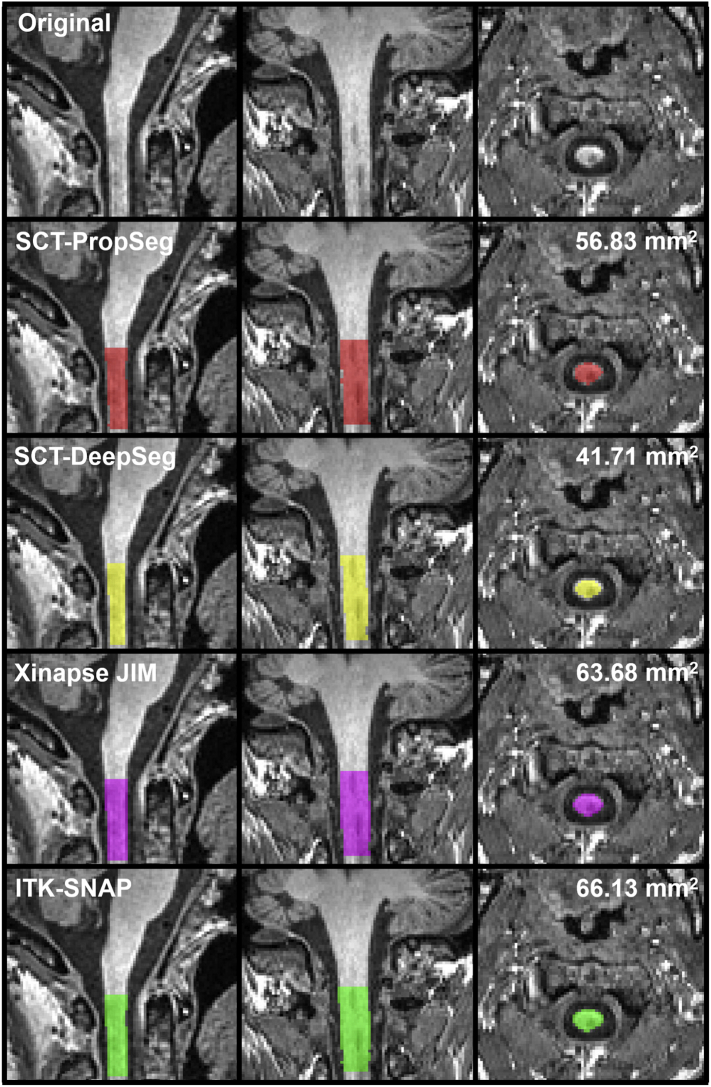
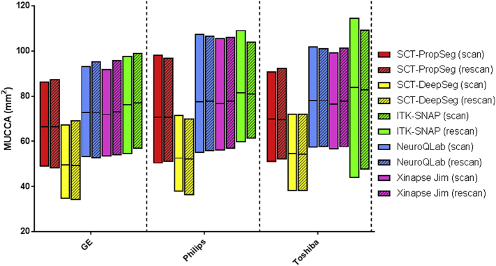
Five methods to measure MUCCA (SCT-PropSeg, SCT-DeepSeg, NeuroQLab, Xinapse JIM and ITK-SNAP) were investigated in a predefined upper cervical cord region. First, within-scanner reproducibility and between-scanner robustness were assessed using intra-class correlation coefficient (ICC) and Dice's similarity index (SI) in scan-rescan 3DT1-weighted images (brain, including cervical spine using a head coil) performed on three 3 T MR machines (GE MR750, Philips Ingenuity, Toshiba Vantage Titan) in 21 subjects with MS and 6 healthy controls (dataset A). Second, sensitivity of MUCCA measurement to lesions in the upper cervical cord was assessed with cervical 3D T1-weighted images (3 T GE HDxT using a head-neck-spine coil) in 7 subjects with MS without and 14 subjects with MS with cervical lesions (dataset B), using ICC and SI with manual reference segmentations.
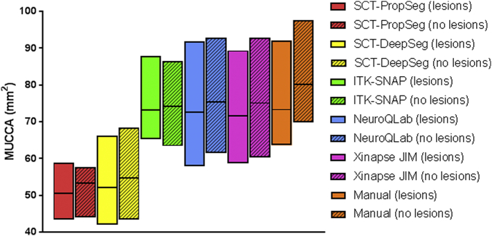
In dataset A, MUCCA differed between MR machines (p < 0.001) and methods (p < 0.001) used, but not between scan sessions. With respect to MUCCA values, Xinapse JIM showed the highest within-scanner reproducibility (ICC absolute agreement = 0.995) while Xinapse JIM and SCT-PropSeg showed the highest between-scanner robustness (ICC consistency = 0.981 and 0.976, respectively). Reproducibility of segmentations between scan sessions was highest in Xinapse JIM and SCT-PropSeg segmentations (median SI ≥ 0.921), with a significant main effect of method (p < 0.001), but not of MR machine or subject group. In dataset B, SI with manual outlines did not differ between patients with or without cervical lesions for any of the segmentation methods (p > 0.176). However, there was an effect of method for both volumetric and voxel wise agreement of the segmentations (both p < 0.001). Highest volumetric and voxel wise agreement was obtained with Xinapse JIM (ICC absolute agreement = 0.940 and median SI = 0.962).
Although MUCCA is highly reproducible within a scanner for each individual measurement method, MUCCA differs between scanners and between methods. Cervical cord lesions do not affect MUCCA measurement performance.
脊髓萎缩已知发生在多发性硬化症(MS)中。可使用平均上颈髓区(MUCCA)来测量这种萎缩。目前,已经存在几种(半)自动化的 MUCCA 测量方法,但在临床磁共振(MR)图像中缺乏验证。
在预定义的上颈髓区域中,研究了 5 种测量 MUCCA 的方法(SCT-PropSeg、SCT-DeepSeg、NeuroQLab、Xinapse JIM 和 ITK-SNAP)。首先,使用受试者内相关系数(ICC)和 Dice 相似性指数(SI),在 21 例 MS 患者和 6 例健康对照者的 3 台 3T MR 机器(GE MR750、Philips Ingenuity、Toshiba Vantage Titan)上的脑扫描-重扫 3DT1 加权图像(包括使用头部线圈的颈椎)中评估了 within-scanner 可重复性和 between-scanner 稳健性(数据集 A)。其次,使用颈部 3D T1 加权图像(使用头部-颈部-脊柱线圈的 3T GE HDxT),在 7 例无颈椎病变的 MS 患者和 14 例有颈椎病变的 MS 患者中评估了 MUCCA 测量对 upper cervical cord 病变的敏感性,使用 ICC 和 SI 与手动参考分割进行评估(数据集 B)。
在数据集 A 中,MR 机器(p<0.001)和使用的方法(p<0.001)之间存在 MUCCA 差异,但扫描会话之间不存在差异。就 MUCCA 值而言,Xinapse JIM 显示出最高的 within-scanner 可重复性(ICC 绝对一致性=0.995),而 Xinapse JIM 和 SCT-PropSeg 显示出最高的 between-scanner 稳健性(ICC 一致性分别为 0.981 和 0.976)。Xinapse JIM 和 SCT-PropSeg 分割的扫描会话之间的分割可重复性最高(中位数 SI≥0.921),方法有显著的主效应(p<0.001),但 MR 机器或受试者组无显著主效应。在数据集 B 中,对于任何分割方法,有或无颈椎病变的患者之间的 SI 均无差异(p>0.176)。然而,分割的体积和体素一致性均存在方法效应(均 p<0.001)。Xinapse JIM 获得了最高的体积和体素一致性(ICC 绝对一致性=0.940,中位数 SI=0.962)。
尽管对于每个单独的测量方法,MUCCA 在扫描仪内的可重复性很高,但 MUCCA 在扫描仪之间和方法之间存在差异。颈椎病变不会影响 MUCCA 测量性能。