Department of Neurology, Dartmouth Hitchcock Medical Center and Geisel School of Medicine, Lebanon, NH, United States.
Front Immunol. 2019 Aug 2;10:1821. doi: 10.3389/fimmu.2019.01821. eCollection 2019.
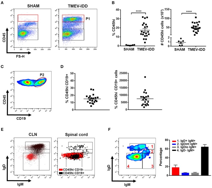
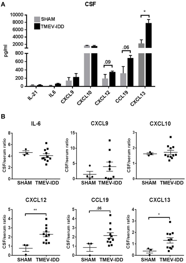
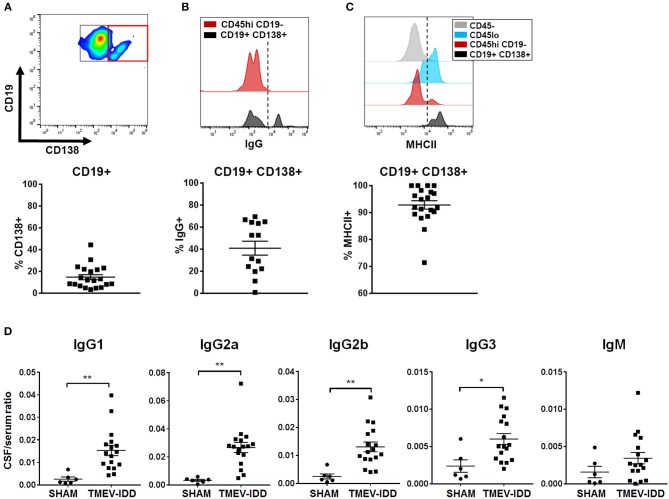
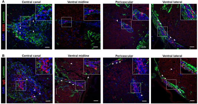
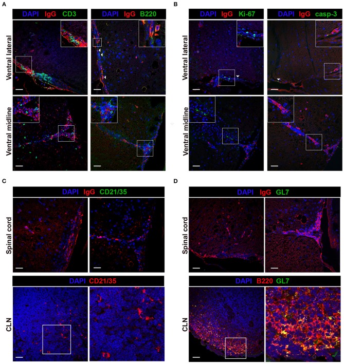
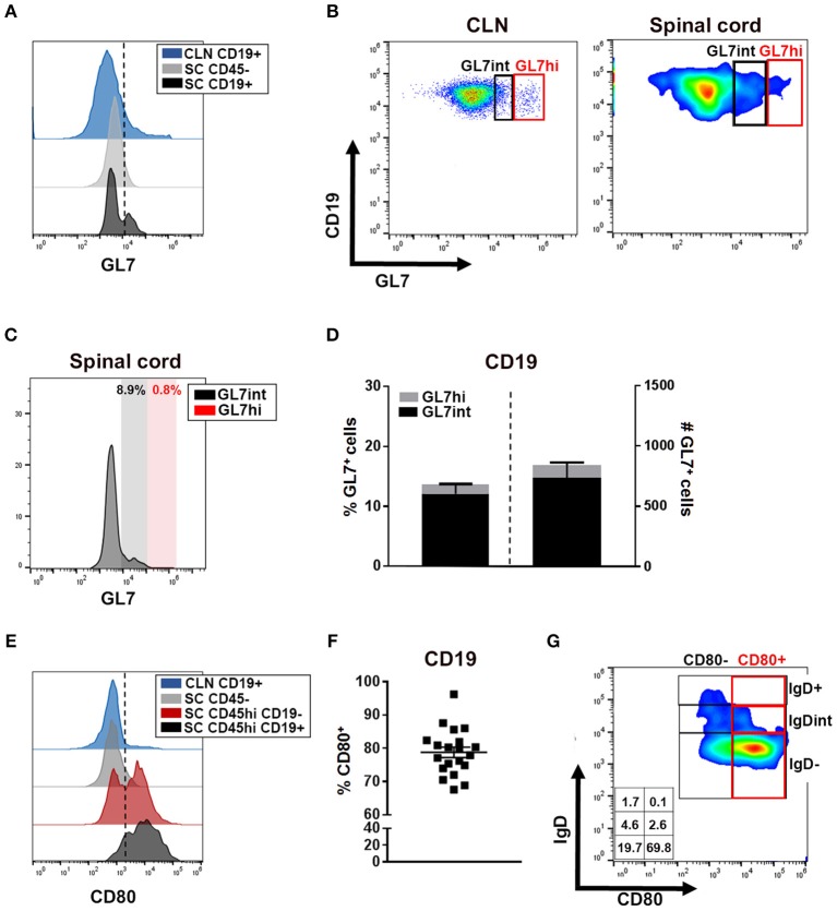
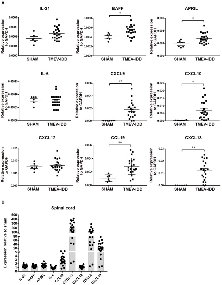
Persistent central nervous system (CNS) inflammation, as seen in chronic infections or inflammatory demyelinating diseases such as Multiple Sclerosis (MS), results in the accumulation of various B cell subsets in the CNS, including naïve, activated, memory B cells (Bmem), and antibody secreting cells (ASC). However, factors driving heterogeneous B cell subset accumulation and antibody (Ab) production in the CNS compartment, including the contribution of ectopic lymphoid follicles (ELF), during chronic CNS inflammation remain unclear and is a major gap in our understanding of neuroinflammation. We sought to address this gap using the Theiler's murine encephalomyelitis virus-induced demyelinating disease (TMEV-IDD) model of progressive MS. In this model, injection of the virus into susceptible mouse strains results in a persistent infection associated with demyelination and progressive disability. During chronic infection, the predominant B cell phenotypes accumulating in the CNS were isotype-switched B cells, including Bmem and ASC with naïve/early activated and transitional B cells present at low frequencies. B cell accumulation in the CNS during chronic TMEV-IDD coincided with intrathecal Ab synthesis in the cerebrospinal fluid (CSF). Mature and isotype-switched B cells predominately localized to the meninges and perivascular space, with IgG isotype-switched B cells frequently accumulating in the parenchymal space. Both mature and isotype-switched B cells and T cells occupied meningeal and perivascular spaces, with minimal evidence for spatial organization typical of ELF mimicking secondary lymphoid organs (SLO). Moreover, immunohistological analysis of immune cell aggregates revealed a lack of SLO-like ELF features, such as cell proliferation, cell death, and germinal center B cell markers. Nonetheless, flow cytometric assessment of B cells within the CNS showed enhanced expression of activation markers, including moderate upregulation of GL7 and expression of the costimulatory molecule CD80. B cell-related chemokines and trophic factors, including APRIL, BAFF, CXCL9, CXCL10, CCL19, and CXCL13, were elevated in the CNS. These results indicate that localization of heterogeneous B cell populations, including activated and isotype-switched B cell phenotypes, to the CNS and intrathecal Ab (ItAb) synthesis can occur independently of SLO-like follicles during chronic inflammatory demyelinating disease.
持续性中枢神经系统(CNS)炎症,如慢性感染或炎症性脱髓鞘疾病(如多发性硬化症[MS])所见,导致各种 B 细胞亚群在 CNS 中积累,包括幼稚、激活、记忆 B 细胞(Bmem)和抗体分泌细胞(ASC)。然而,导致慢性 CNS 炎症时 CNS 区室中异质性 B 细胞亚群积累和抗体(Ab)产生的因素,包括异位淋巴滤泡(ELF)的贡献,仍不清楚,这是我们对神经炎症理解的主要空白。我们试图使用实验性自身免疫性脑脊髓炎病毒诱导的脱髓鞘疾病(EAE)模型来解决这一空白,这是一种进展性 MS 的多发性硬化症模型。在该模型中,将病毒注射到易感小鼠株中会导致持续性感染,从而导致脱髓鞘和进行性残疾。在慢性感染期间,在 CNS 中积累的主要 B 细胞表型是同种型转换 B 细胞,包括 Bmem 和 ASC,幼稚/早期激活和过渡 B 细胞的频率较低。慢性 TMEV-IDD 时 CNS 中的 B 细胞积累与脑脊液(CSF)中的鞘内 Ab 合成一致。成熟和同种型转换的 B 细胞主要定位于脑膜和血管周围空间,IgG 同种型转换的 B 细胞经常在实质空间中积累。成熟和同种型转换的 B 细胞和 T 细胞占据脑膜和血管周围空间,很少有证据表明存在类似于次级淋巴器官(SLO)的空间组织。此外,免疫细胞聚集体的免疫组织化学分析显示缺乏 SLO 样 ELF 特征,例如细胞增殖、细胞死亡和生发中心 B 细胞标志物。尽管如此,对 CNS 内 B 细胞的流式细胞术评估显示激活标志物表达增强,包括 GL7 的中度上调和共刺激分子 CD80 的表达。B 细胞相关趋化因子和营养因子,包括 APRIL、BAFF、CXCL9、CXCL10、CCL19 和 CXCL13,在 CNS 中升高。这些结果表明,包括激活和同种型转换的 B 细胞表型在内的异质性 B 细胞群体向 CNS 的定位和鞘内 Ab(ItAb)合成可以在慢性炎症性脱髓鞘疾病期间独立于 SLO 样滤泡发生。