Kaneko Junichi, Matsubayashi Hiroyuki, Satoh Tatsunori, Sato Junya, Takinami Masaki, Ishiwatari Hirotoshi, Uesaka Katsuhiko, Abe Masato, Sasaki Keiko, Ono Hiroyuki
Division of Endoscopy, Shizuoka Cancer Center, Japan.
Division of Hepato-pancreaticobiliary Surgery, Shizuoka Cancer Center, Japan.
Intern Med. 2020 Jan 15;59(2):199-204. doi: 10.2169/internalmedicine.3561-19. Epub 2019 Sep 3.
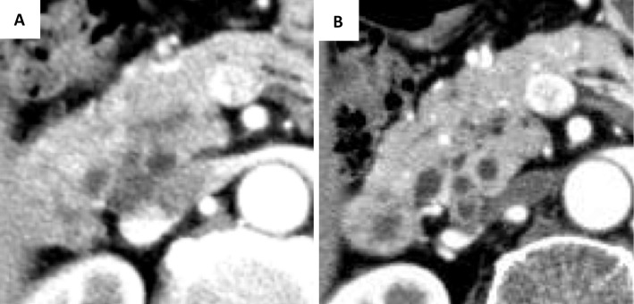
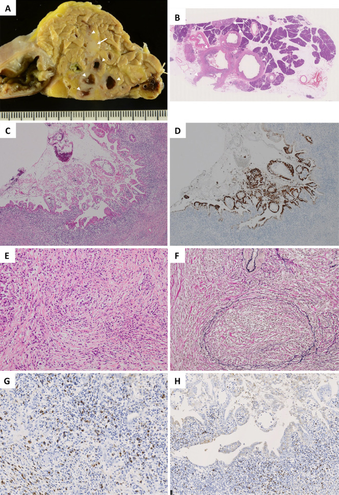
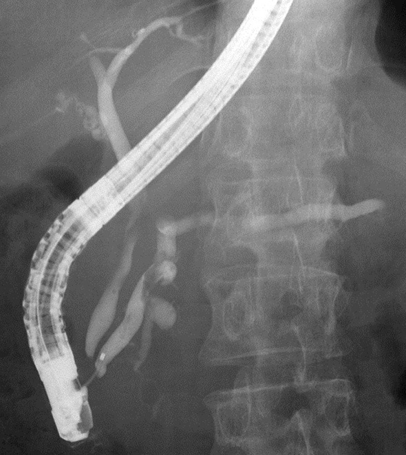
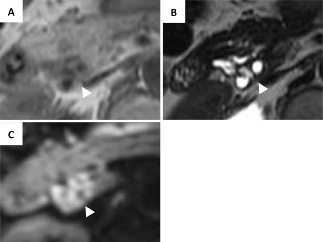
A small proportion of intraductal papillary mucinous neoplasms (IPMNs) are accompanied by type 1 autoimmune pancreatitis (AIP); however their clinical courses and image characteristics have not been fully reported. A 65-year-old woman was referred to our hospital for the examination of a pancreatic head cyst that had shown exacerbation for two years. Several images demonstrated a multilocular cyst with a symmetrically thickened, enhanced, cyst wall. Cancerization of IPMN was suspected, and pancreatoduodenectomy was performed. The resected specimens showed a multilocular cyst with solid areas. The solid areas demonstrated pathological findings that corresponded with type 1 AIP. Papillary epithelia suggestive of IPMN was recognized in some parts of the cystic wall.
一小部分导管内乳头状黏液性肿瘤(IPMN)伴有1型自身免疫性胰腺炎(AIP);然而,它们的临床病程和影像特征尚未得到充分报道。一名65岁女性因胰头囊肿检查被转诊至我院,该囊肿已加重两年。多项影像显示为多房囊肿,囊壁对称增厚、强化。怀疑IPMN癌变,遂行胰十二指肠切除术。切除标本显示为带有实性区域的多房囊肿。实性区域显示出与1型AIP相符的病理结果。在囊壁的某些部位可识别出提示IPMN的乳头状上皮。