Murakami Yuryo, Ueki Ryusuke, Tachikawa Taihei, Hirose Munetaka
Department of Anesthesiology and Pain Medicine, Hyogo College of Medicine, Nishinomiya, Japan.
Department of Anesthesiology, Meiwa Hospital, Nishinomiya, Japan.
Anesth Pain Med. 2019 Apr 23;9(3):e89417. doi: 10.5812/aapm.89417. eCollection 2019 Jun.
The pathophysiological mechanism of propofol-related infusion syndrome (PRIS) is believed to be due to the injury to the mitochondrial electron transport chain and the resultant metabolic disorders that are caused by both propofol agents and the lipid solvent. However, the mechanisms and causative factors of PRIS have not been fully elucidated.
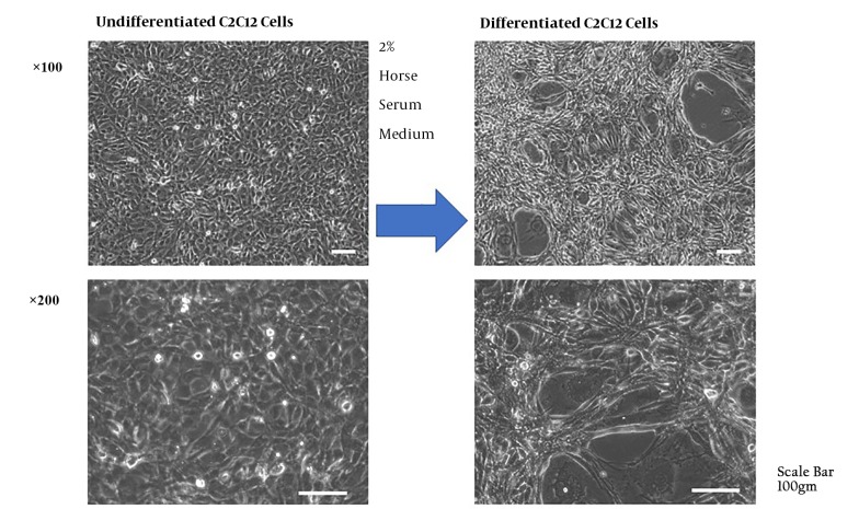
The aim of this study was to evaluate the possibility of a research model using the culture of differentiated C2C12 cells for fundamental research of PRIS.
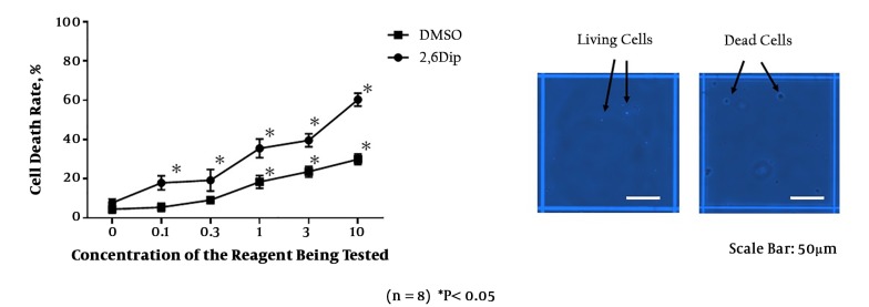
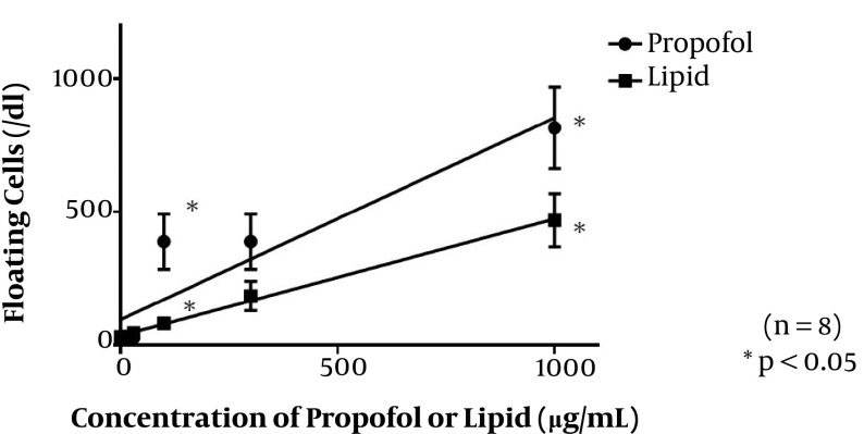
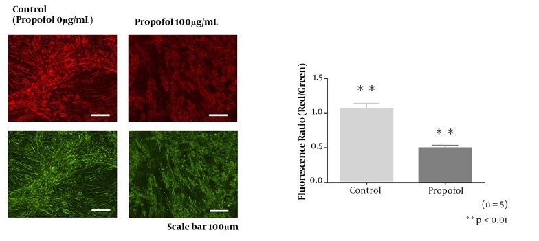
First, differentiated C2C12 cells were cultured accompanied by several concentrations of chemical reagents of 2,6-diisopropylphenol (2,6 DIP) or dimethyl sulfoxide (DMSO) for 60 hours and the cell death rate was examined by trypan blue staining. Second, The cells were incubated with a commercially available propofol reagent or lipid reagent for 48 hours. The supernatant fluid of the cell culture medium was gathered and the numbers of floating cells were measured by cell counter. To investigate the mitochondrial disorder by the propofol preparation, JC-1, an experiment using fluorescent reagent, was performed for the 48 hours with 100 µg/mL propofol incubation.
The rate of cell death was increased with elevating concentrations both of chemical reagents of 2,6 DIP group and dimethyl sulfoxide group. The rates of cell death were significantly higher in the 2,6 DIP group than DMSO group. The numbers of floating cells were increased with elevating concentrations both commercially available propofol reagent and lipid reagent groups. The decreased red/green fluorescence ratio by JC-1 staining in the propofol 100µg/mL group proved an attenuated mitochondrial membrane potential.
The dose-dependent cell damage induced by the propofol reagents and a lipid solvent may provide a proposed model as a basic experimental model for further investigations into PRIS.
丙泊酚相关输注综合征(PRIS)的病理生理机制被认为是由于丙泊酚制剂和脂质溶剂对线粒体电子传递链的损伤以及由此导致的代谢紊乱。然而,PRIS的机制和致病因素尚未完全阐明。
本研究的目的是评估使用分化的C2C12细胞培养物作为PRIS基础研究的研究模型的可能性。
首先,将分化的C2C12细胞与几种浓度的2,6-二异丙基苯酚(2,6-DIP)或二甲基亚砜(DMSO)化学试剂一起培养60小时,并用台盼蓝染色检测细胞死亡率。其次,将细胞与市售丙泊酚试剂或脂质试剂孵育48小时。收集细胞培养基的上清液,并用细胞计数器测量漂浮细胞的数量。为了研究丙泊酚制剂引起的线粒体紊乱,使用荧光试剂JC-1进行实验,用100μg/mL丙泊酚孵育48小时。
2,6-DIP组和二甲基亚砜组化学试剂浓度升高时,细胞死亡率均升高。2,6-DIP组的细胞死亡率明显高于DMSO组。市售丙泊酚试剂组和脂质试剂组中,漂浮细胞数量均随浓度升高而增加。丙泊酚100μg/mL组中JC-1染色导致的红/绿荧光比值降低证明线粒体膜电位减弱。
丙泊酚试剂和脂质溶剂诱导的剂量依赖性细胞损伤可能为进一步研究PRIS提供一个作为基础实验模型的建议模型。