Department of Anatomy, Histology and Embryology, Shanghai Medical School of Fudan University, 138 Yixueyuan Road, Shanghai, 200032, People's Republic of China.
Basic Res Cardiol. 2019 Oct 6;114(6):43. doi: 10.1007/s00395-019-0752-z.
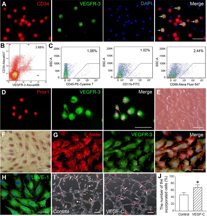
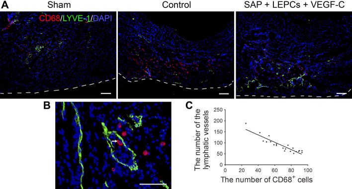
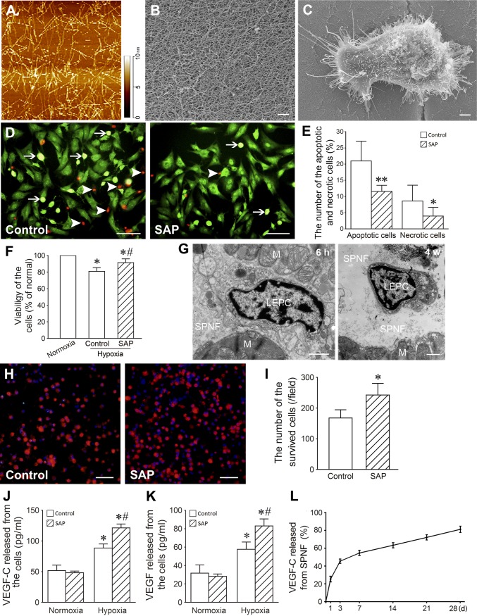
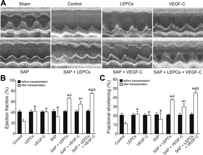
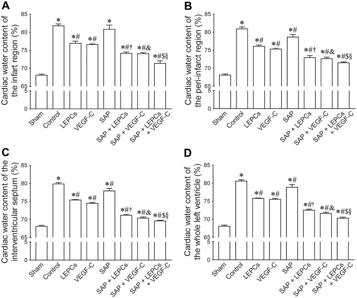
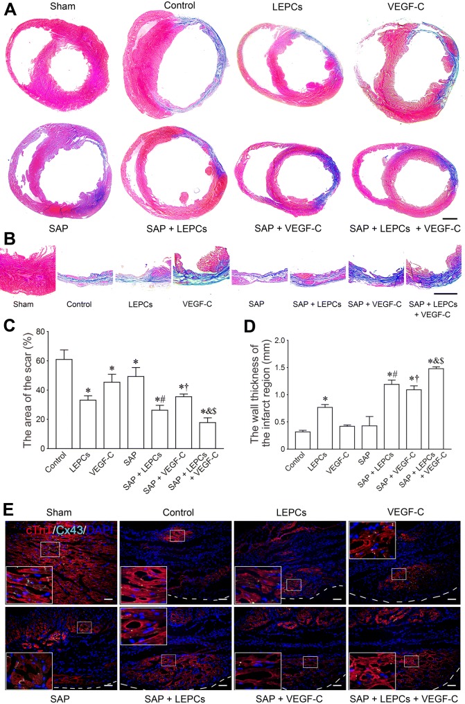
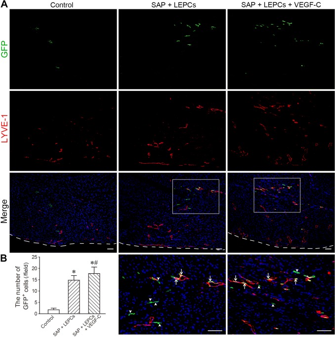
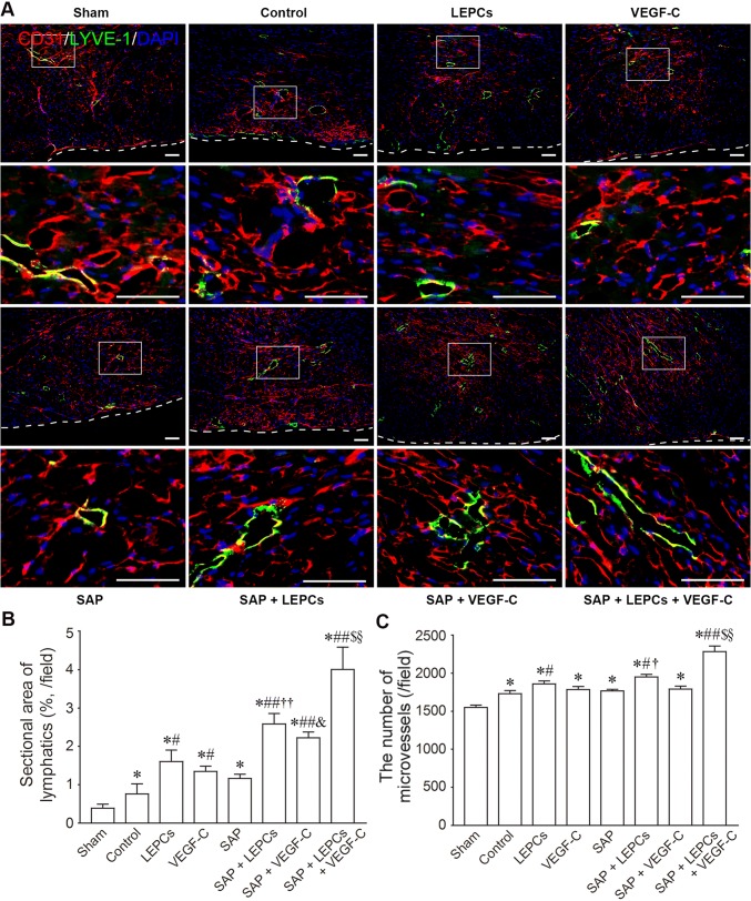
Impairment of cardiac lymphatic vessels leads to cardiac lymphedema. Recent studies have suggested that stimulation of lymphangiogenesis may reduce cardiac lymphedema. However, effects of lymphatic endothelial progenitor cells (LEPCs) on cardiac lymphangiogenesis are poorly understood. Therefore, this study investigated effectiveness of LEPC transplantation and VEGF-C release with self-assembling peptide (SAP) on cardiac lymphangiogenesis after myocardial infarction (MI). CD34VEGFR-3 EPCs isolated from rat bone marrow differentiated into lymphatic endothelial cells after VEGF-C induction. VEGF-C also stimulated the cells to incorporate into the lymphatic capillary-like structures. The functionalized SAP could adhere with the cells and released VEGF-C sustainedly. In the condition of hypoxia and serum deprivation or abdominal pouch assay, the SAP hydrogel protected the cells from apoptosis and necrosis. At 4 weeks after intramyocardial transplantation of the cells and VEGF-C loaded with SAP hydrogel in rat MI models, cardiac lymphangiogenesis was increased, cardiac edema and reverse remodeling were reduced, and cardiac function was improved significantly. Delivery with SAP hydrogel favored survival of the engrafted cells. VEGF-C released from the hydrogel promoted differentiation and incorporation of the cells as well as growth of pre-existed lymphatic vessels. Cardiac lymphangiogenesis was beneficial for elimination of the inflammatory cells in the infarcted myocardium. Moreover, angiogenesis and myocardial regeneration were enhanced after reduction of lymphedema. These results demonstrate that the combined delivery of LEPCs and VEGF-C with the functionalized SAP promotes cardiac lymphangiogenesis and repair of the infarcted myocardium effectively. This study represents a novel therapy for relieving myocardial edema in cardiovascular diseases.
心脏淋巴管的损伤会导致心脏淋巴水肿。最近的研究表明,刺激淋巴管生成可能会减少心脏淋巴水肿。然而,淋巴管内皮祖细胞(LEPCs)对心脏淋巴管生成的影响还知之甚少。因此,本研究探讨了 LEPC 移植和自组装肽(SAP)释放 VEGF-C 对心肌梗死后(MI)心脏淋巴管生成的有效性。从大鼠骨髓中分离的 CD34VEGFR-3 EPC 在 VEGF-C 诱导下分化为淋巴管内皮细胞。VEGF-C 还刺激细胞形成淋巴管样毛细血管结构。功能化的 SAP 可以与细胞结合,并持续释放 VEGF-C。在缺氧和血清剥夺或腹部囊检测条件下,SAP 水凝胶可防止细胞凋亡和坏死。在大鼠 MI 模型中,将细胞和 SAP 水凝胶负载的 VEGF-C 经心肌内移植 4 周后,心脏淋巴管生成增加,心脏水肿和逆重构减少,心功能明显改善。SAP 水凝胶的递送有利于移植细胞的存活。水凝胶中释放的 VEGF-C 促进了细胞的分化和整合以及预先存在的淋巴管的生长。心脏淋巴管生成有利于清除梗死心肌中的炎症细胞。此外,淋巴管生成和心肌再生在减轻淋巴水肿后得到增强。这些结果表明,LEPCs 和 VEGF-C 与功能化 SAP 的联合递送可有效促进心脏淋巴管生成和梗死心肌的修复。本研究为减轻心血管疾病中心肌水肿提供了一种新的治疗方法。