Department of Gastroenterology, Justus Liebig University, D-35392 Giessen, Germany.
Max Planck Institute for Heart and Lung Research, Member of the German Center for Lung Research (DZL), Member of the Cardio-Pulmonary Institute (CPI), D-61231 Bad Nauheim, Germany.
Cells. 2019 Oct 7;8(10):1210. doi: 10.3390/cells8101210.
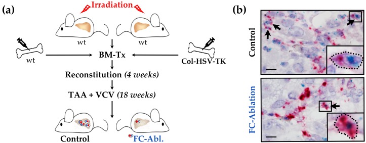
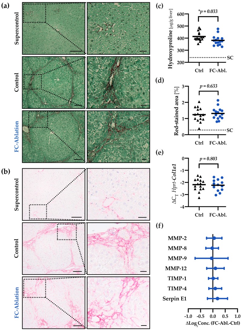
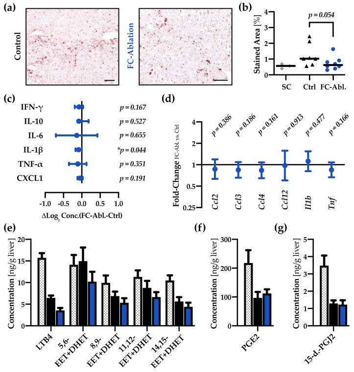
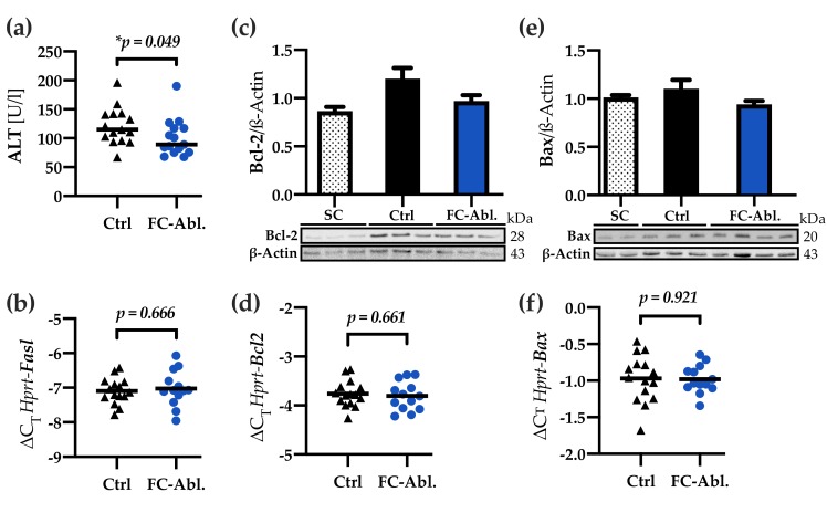
Bone marrow-derived fibrocytes (FC) represent a unique cell type, sharing features of both mesenchymal and hematopoietic cells. FC were shown to specifically infiltrate the injured liver and participate in fibrogenesis. Moreover, FC exert a variety of paracrine functions, thus possibly influencing the disease progression. However, the overall contribution of FC to liver fibrosis remains unclear. We aimed to study the effect of a specific FC depletion, utilizing a herpes simplex virus thymidine kinase (HSV-TK)/Valganciclovir suicide gene strategy. Fibrosis was induced by oral thioacetamide (TAA) administration in C57BL/6J mice. Hepatic hydroxyproline content was assessed for the primary readout. The HSV-TK model enabled the specific depletion of fibrocytes. Hepatic hydroxyproline content was significantly reduced as a result of the fibrocyte ablation (-7.8%; 95% CI: 0.7-14.8%; = 0.033), denoting a reduced deposition of fibrillar collagens. Lower serum alanine transaminase levels (-20.9%; 95% CI: 0.4-36.9%; = 0.049) indicate a mitigation of liver-specific cellular damage. A detailed mode of action, however, remains yet to be identified. The present study demonstrates a relevant functional contribution of fibrocytes to chronic toxic liver fibrosis, contradicting recent reports. Our results emphasize the need to thoroughly study the biology of fibrocytes in order to understand their importance for hepatic fibrogenesis.
骨髓来源的成纤维细胞 (FC) 代表了一种独特的细胞类型,具有间充质和造血细胞的特征。FC 被证明会特异性浸润受损的肝脏并参与纤维化形成。此外,FC 发挥多种旁分泌功能,从而可能影响疾病进展。然而,FC 对肝纤维化的总体贡献仍不清楚。我们旨在利用单纯疱疹病毒胸苷激酶 (HSV-TK)/缬更昔洛韦自杀基因策略,研究特异性耗尽 FC 的效果。通过给予 C57BL/6J 小鼠口服硫代乙酰胺 (TAA) 来诱导纤维化。羟脯氨酸含量评估作为主要的研究结果。HSV-TK 模型可实现成纤维细胞的特异性耗竭。由于 FC 消融,肝羟脯氨酸含量显著降低(-7.8%;95%CI:0.7-14.8%; = 0.033),表示纤维胶原的沉积减少。较低的血清丙氨酸氨基转移酶水平(-20.9%;95%CI:0.4-36.9%; = 0.049)表明肝脏特异性细胞损伤减轻。然而,详细的作用机制仍有待确定。本研究表明 FC 对慢性毒性肝纤维化具有相关的功能贡献,这与最近的报告相矛盾。我们的结果强调需要深入研究 FC 的生物学特性,以了解它们对肝纤维化形成的重要性。