Institute of Structural and Molecular Biology, Birkbeck, University of London, Malet Street, London, United Kingdom.
Institute of Structural and Molecular Biology, Birkbeck, University of London, Malet Street, London, United Kingdom.
J Struct Biol. 2020 Jan 1;209(1):107402. doi: 10.1016/j.jsb.2019.10.004. Epub 2019 Oct 11.
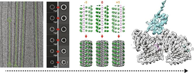
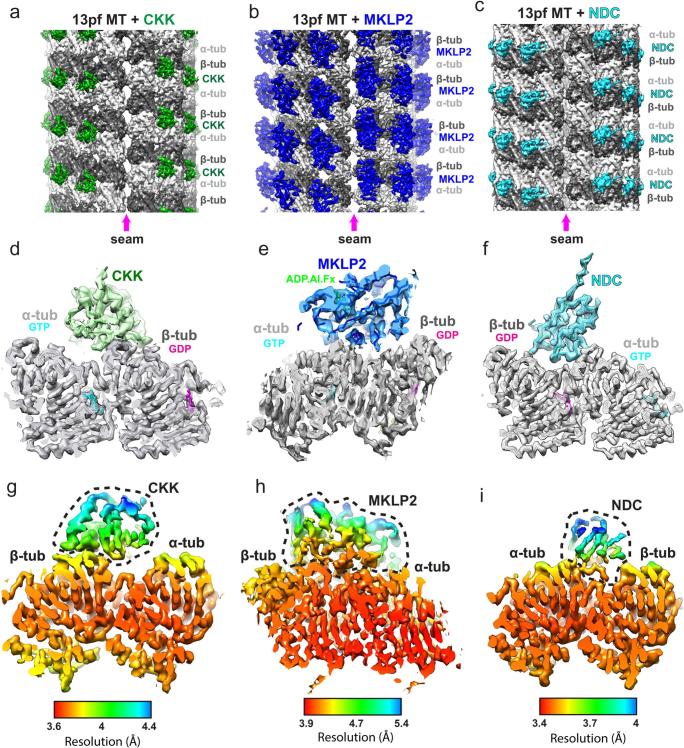
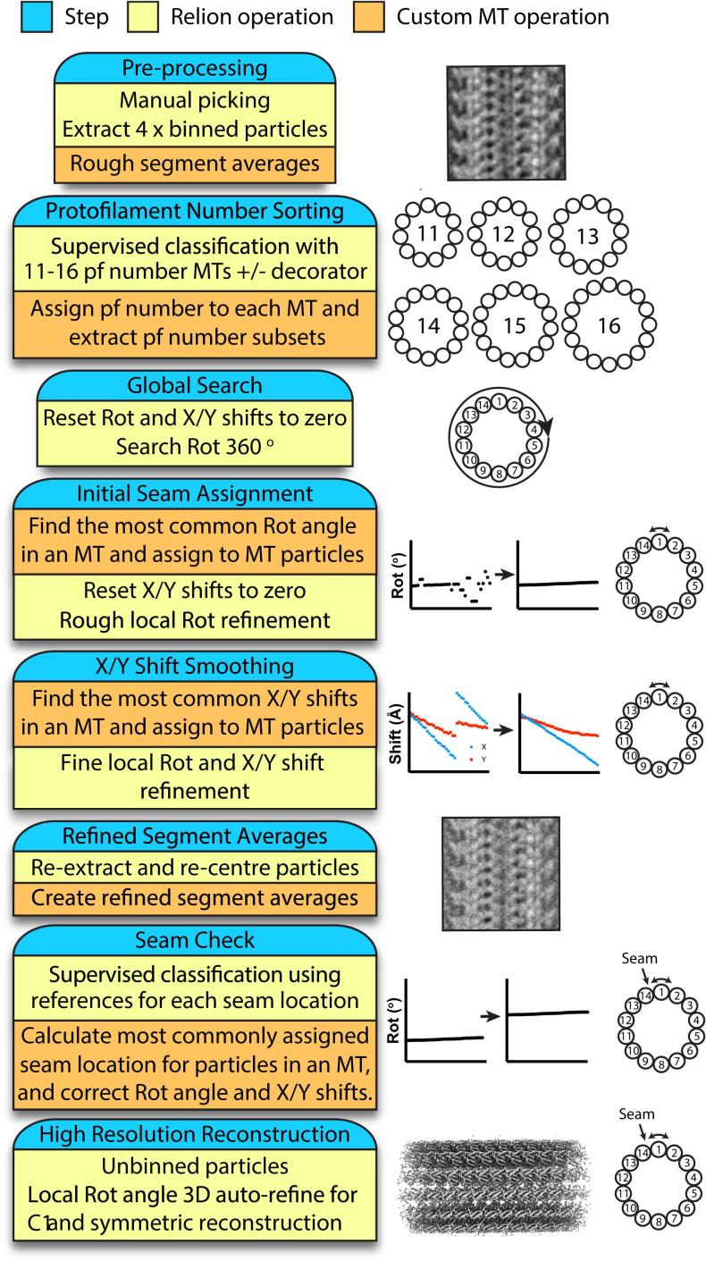
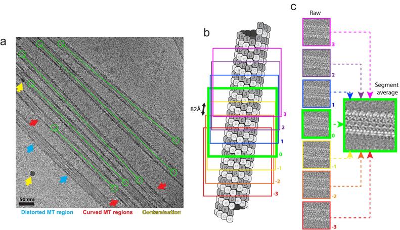
Microtubules are polar filaments built from αβ-tubulin heterodimers that exhibit a range of architectures in vitro and in vivo. Tubulin heterodimers are arranged helically in the microtubule wall but many physiologically relevant architectures exhibit a break in helical symmetry known as the seam. Noisy 2D cryo-electron microscopy projection images of pseudo-helical microtubules therefore depict distinct but highly similar views owing to the high structural similarity of α- and β-tubulin. The determination of the αβ-tubulin register and seam location during image processing is essential for alignment accuracy that enables determination of biologically relevant structures. Here we present a pipeline designed for image processing and high-resolution reconstruction of cryo-electron microscopy microtubule datasets, based in the popular and user-friendly RELION image-processing package, Microtubule RELION-based Pipeline (MiRP). The pipeline uses a combination of supervised classification and prior knowledge about geometric lattice constraints in microtubules to accurately determine microtubule architecture and seam location. The presented method is fast and semi-automated, producing near-atomic resolution reconstructions with test datasets that contain a range of microtubule architectures and binding proteins.
微管是由αβ-微管蛋白异二聚体构成的极性细丝,在体外和体内表现出多种结构。微管蛋白异二聚体在微管壁中呈螺旋排列,但许多与生理相关的结构表现出螺旋对称的断裂,称为 seam。由于α-和β-微管蛋白具有很高的结构相似性,因此伪螺旋微管的嘈杂 2D 冷冻电子显微镜投影图像描绘了独特但高度相似的视图。因此,在图像处理过程中确定αβ-微管蛋白的登记和 seam 位置对于对齐精度至关重要,这可以确定与生物学相关的结构。在这里,我们提出了一个基于流行且用户友好的 RELION 图像处理包的图像处理和冷冻电子显微镜微管数据集高分辨率重建的流水线,称为基于 RELION 的微管图像处理流水线 (MiRP)。该流水线使用监督分类和微管中几何晶格约束的先验知识的组合,准确地确定微管结构和 seam 位置。所提出的方法快速且半自动,使用包含多种微管结构和结合蛋白的测试数据集生成近原子分辨率的重建。