Department of Radiology, Washington University School of Medicine, St. Louis, MO, USA.
Department of Medicine, Division of Oncology, Washington University School of Medicine, St. Louis, MO, USA.
Eur J Nucl Med Mol Imaging. 2022 Jan;49(2):550-562. doi: 10.1007/s00259-021-05489-8. Epub 2021 Jul 30.
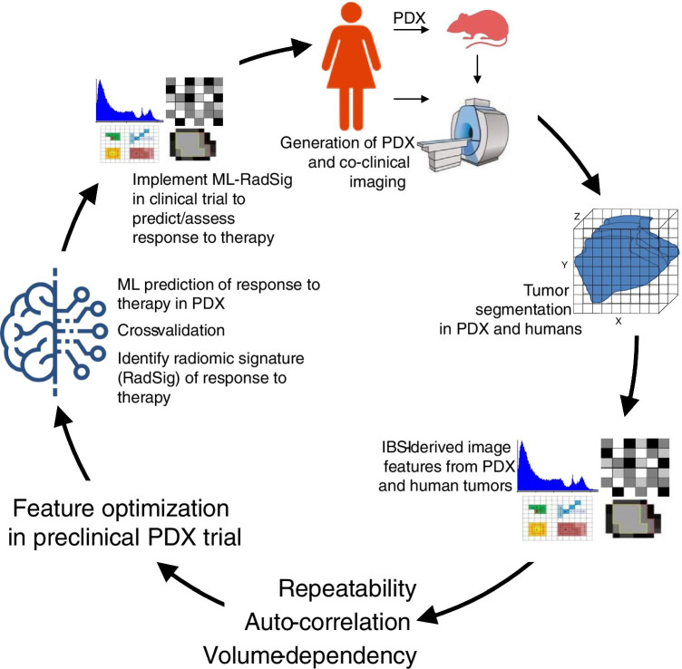
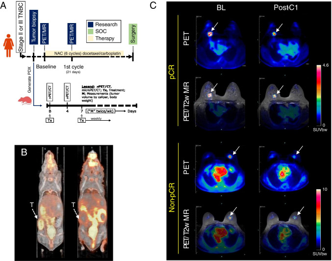
We sought to exploit the heterogeneity afforded by patient-derived tumor xenografts (PDX) to first, optimize and identify robust radiomic features to predict response to therapy in subtype-matched triple negative breast cancer (TNBC) PDX, and second, to implement PDX-optimized image features in a TNBC co-clinical study to predict response to therapy using machine learning (ML) algorithms.
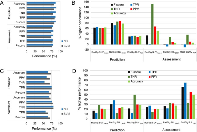
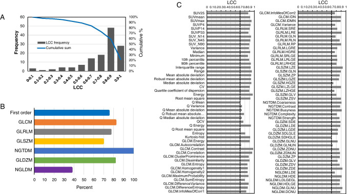
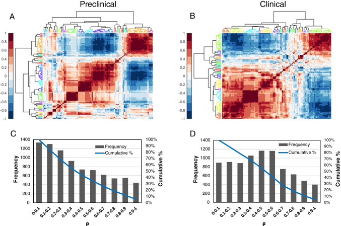
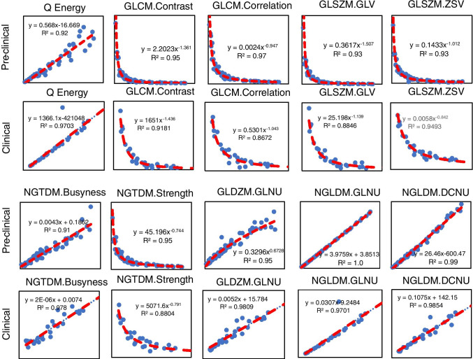
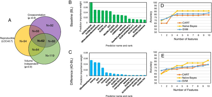
TNBC patients and subtype-matched PDX were recruited into a co-clinical FDG-PET imaging trial to predict response to therapy. One hundred thirty-one imaging features were extracted from PDX and human-segmented tumors. Robust image features were identified based on reproducibility, cross-correlation, and volume independence. A rank importance of predictors using ReliefF was used to identify predictive radiomic features in the preclinical PDX trial in conjunction with ML algorithms: classification and regression tree (CART), Naïve Bayes (NB), and support vector machines (SVM). The top four PDX-optimized image features, defined as radiomic signatures (RadSig), from each task were then used to predict or assess response to therapy. Performance of RadSig in predicting/assessing response was compared to SUV, SUV, and lean body mass-normalized SUL measures.
Sixty-four out of 131 preclinical imaging features were identified as robust. NB-RadSig performed highest in predicting and assessing response to therapy in the preclinical PDX trial. In the clinical study, the performance of SVM-RadSig and NB-RadSig to predict and assess response was practically identical and superior to SUV, SUV, and SUL measures.
We optimized robust FDG-PET radiomic signatures (RadSig) to predict and assess response to therapy in the context of a co-clinical imaging trial.
我们试图利用患者来源的肿瘤异种移植(PDX)的异质性,首先优化并确定稳健的放射组学特征,以预测三阴性乳腺癌(TNBC)PDX 中治疗的反应,其次,将 PDX 优化的图像特征应用于 TNBC 联合临床研究中,使用机器学习(ML)算法预测治疗反应。
招募 TNBC 患者和匹配亚型的 PDX 参加 FDG-PET 联合临床成像试验,以预测治疗反应。从 PDX 和人体分割肿瘤中提取了 131 个成像特征。基于可重复性、互相关和体积独立性,确定了稳健的图像特征。使用 ReliefF 对预测因子的排名重要性进行分析,以结合 ML 算法(分类和回归树(CART)、朴素贝叶斯(NB)和支持向量机(SVM))在临床前 PDX 试验中识别预测性放射组学特征。然后,使用来自每个任务的前四个 PDX 优化的图像特征(定义为放射组学特征(RadSig))来预测或评估治疗反应。将 RadSig 预测/评估反应的性能与 SUV、SUV 和瘦体重归一化 SUL 测量值进行比较。
在 131 个临床前成像特征中,有 64 个被确定为稳健。NB-RadSig 在预测和评估临床前 PDX 试验中的治疗反应方面表现最佳。在临床研究中,SVM-RadSig 和 NB-RadSig 预测和评估反应的性能几乎相同,优于 SUV、SUV 和 SUL 测量值。
我们优化了稳健的 FDG-PET 放射组学特征(RadSig),以在联合临床成像试验中预测和评估治疗反应。