Center for Modeling, Simulation and Imaging in Medicine (CeMSIM), Rensselaer Polytechnic Institute, Troy, NY, USA.
The Department of Chemical and Biological Engineering, Rensselaer Polytechnic Institute, Troy, NY, USA.
Sci Rep. 2019 Dec 16;9(1):19138. doi: 10.1038/s41598-019-55012-1.
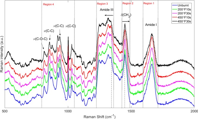
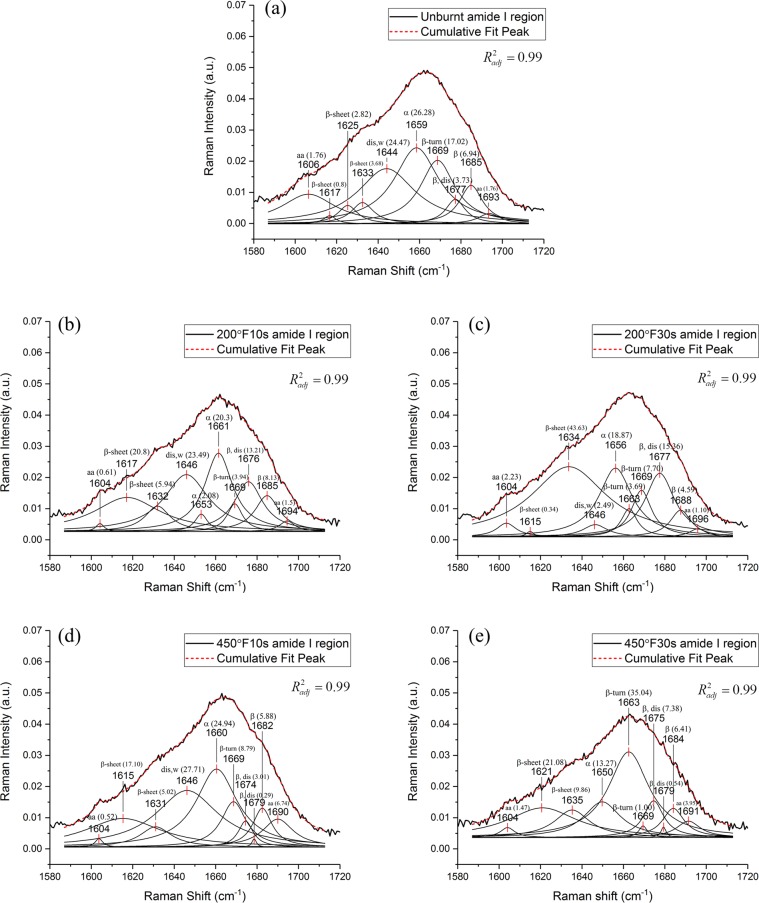
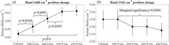
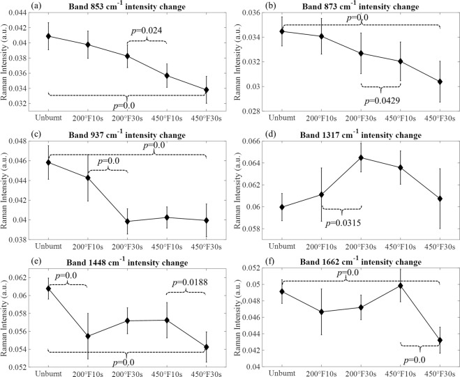
This study utilizes Raman spectroscopy to analyze the burn-induced collagen conformational changes in ex vivo porcine skin tissue. Raman spectra of wavenumbers 500-2000 cm were measured for unburnt skin as well as four different burn conditions: (i) 200 °F for 10 s, (ii) 200 °F for the 30 s, (iii) 450 °F for 10 s and (iv) 450 °F for 30 s. The overall spectra reveal that protein and amino acids-related bands have manifested structural changes including the destruction of protein-related functional groups, and transformation from α-helical to disordered structures which are correlated with increasing burn severity. The deconvolution of the amide I region (1580-1720 cm) and the analysis of the sub-bands reveal a change of the secondary structure of the collagen from the α-like helix dominated to the β-aggregate dominated one. Such conformational changes may explain the softening of mechanical response in burnt tissues reported in the literature.
本研究利用拉曼光谱分析了离体猪皮组织烧伤诱导的胶原构象变化。对未烧伤皮肤以及四种不同烧伤情况(i)200°F 10 秒,(ii)200°F 30 秒,(iii)450°F 10 秒和(iv)450°F 30 秒的皮肤分别测量了波数为 500-2000cm 的拉曼光谱。整体光谱表明,与烧伤严重程度增加相关的蛋白和氨基酸相关带已经表现出结构变化,包括破坏蛋白相关功能基团,以及从α-螺旋结构向无序结构的转变。酰胺 I 区域(1580-1720cm)的解卷积和子带分析表明,胶原的二级结构从以α-螺旋为主转变为以β-聚集为主。这种构象变化可能解释了文献中报道的烧伤组织力学响应软化的原因。