Biomedicine Discovery Institute, Department of Anatomy and Developmental Biology, Monash University, Melbourne, Australia.
Sci Rep. 2019 Dec 27;9(1):19941. doi: 10.1038/s41598-019-56423-w.
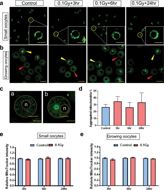
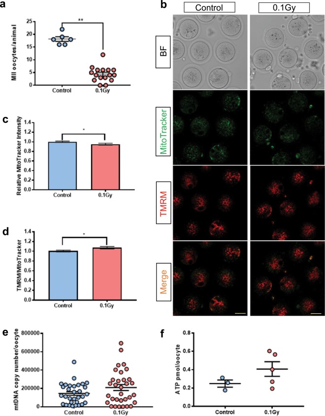
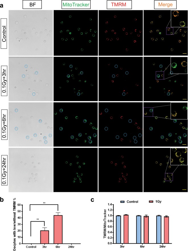
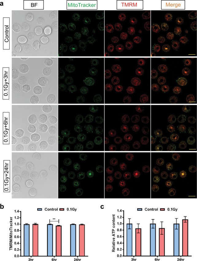
Standard cytotoxic cancer treatments, such as radiation, can damage and deplete the supply of oocytes stored within the ovary, which predisposes females to infertility and premature menopause later in life. The mechanisms by which radiation induces oocyte damage have not been completely elucidated. The objective of this study was to determine if γ-irradiation changes mitochondrial characteristics in oocytes, possibly contributing to a reduction in oocyte number and quality. Immature oocytes were collected from postnatal day (PN) 9-11 C57Bl6 mice 3, 6 and 24 hours after 0.1 Gy γ-irradiation to monitor acute mitochondrial changes. Oocytes were classified as small (>20 µm) or growing (40-60 µm). Mitochondrial membrane potential was lost in 20% and 44% of small oocytes (~20 µm) at 3 and 6 hours after γ-irradiation, respectively, consistent with the induction of apoptosis. However, mitochondrial mass, distribution and membrane potential in the surviving small oocytes were similar to the non-irradiated controls at both time points. At 24 hours after γ-irradiation, all mitochondrial parameters analysed within immature oocytes were similar to untreated controls. Mitochondrial parameters within growing oocytes were also similar to untreated controls. When mice were superovulated more than 3 weeks after γ-irradiation, there was a significant reduction in the number of mature oocytes harvested compared to controls (Control 18 ± 1 vs 0.1 Gy 4 ± 1, n = 6/16 mice, p < 0.05). There was a slight reduction in mitochondrial mass in mature oocytes after γ-irradiation, though mitochondrial localization, mtDNA copy number and ATP levels were similar between groups. In summary, this study shows that γ-irradiation of pre-pubertal mice is associated with loss of mitochondrial membrane potential in a significant proportion of small immature oocytes and a reduction in the number of mature oocytes harvested from adult mice. Furthermore, these results suggest that immature oocytes that survive γ-irradiation and develop through to ovulation contain mitochondria with normal characteristics. Whether the oocytes that survive radiation and eventually undergo meiosis can support fertility remains to be determined.
标准的细胞毒性癌症治疗方法,如放疗,会损害和耗尽卵巢中储存的卵母细胞供应,使女性易患不孕和过早绝经。放疗诱导卵母细胞损伤的机制尚未完全阐明。本研究旨在确定 γ 射线照射是否会改变卵母细胞的线粒体特征,从而导致卵母细胞数量和质量减少。从小鼠出生后第 9-11 天(PN)收集未成熟卵母细胞,在 0.1Gy γ 射线照射后 3、6 和 24 小时监测急性线粒体变化。卵母细胞分为小卵母细胞(>20µm)和生长卵母细胞(40-60µm)。在 3 和 6 小时后,分别有 20%和 44%的小卵母细胞(~20µm)失去线粒体膜电位,这与凋亡的诱导一致。然而,在这两个时间点,存活的小卵母细胞中的线粒体质量、分布和膜电位与未照射对照组相似。在 γ 射线照射后 24 小时,所有分析的未成熟卵母细胞中的线粒体参数均与未处理对照组相似。生长卵母细胞中的线粒体参数也与未处理对照组相似。当小鼠在 γ 射线照射后超过 3 周进行超排卵时,与对照组相比,收获的成熟卵母细胞数量显著减少(对照组 18±1 与 0.1Gy 4±1,n=6/16 只小鼠,p<0.05)。γ 射线照射后成熟卵母细胞中线粒体质量略有减少,但两组间线粒体定位、mtDNA 拷贝数和 ATP 水平相似。总之,本研究表明,对青春期前小鼠进行 γ 射线照射会导致大量小未成熟卵母细胞中线粒体膜电位丧失,并导致从成年小鼠中收获的成熟卵母细胞数量减少。此外,这些结果表明,在 γ 射线照射后存活并发育到排卵的未成熟卵母细胞中含有具有正常特征的线粒体。存活的辐射卵母细胞最终能否进行减数分裂以支持生育能力仍有待确定。