• 文献检索
  • 文档翻译
  • 深度研究
  • 学术资讯
  • Suppr Zotero 插件Zotero 插件
  • 邀请有礼
  • 套餐&价格
  • 历史记录
应用&插件
Suppr Zotero 插件Zotero 插件浏览器插件Mac 客户端Windows 客户端微信小程序
定价
高级版会员购买积分包购买API积分包
服务
文献检索文档翻译深度研究API 文档MCP 服务
关于我们
关于 Suppr公司介绍联系我们用户协议隐私条款
关注我们

Suppr 超能文献

核心技术专利:CN118964589B侵权必究
粤ICP备2023148730 号-1Suppr @ 2026

文献检索

告别复杂PubMed语法,用中文像聊天一样搜索,搜遍4000万医学文献。AI智能推荐,让科研检索更轻松。

立即免费搜索

文件翻译

保留排版,准确专业,支持PDF/Word/PPT等文件格式,支持 12+语言互译。

免费翻译文档

深度研究

AI帮你快速写综述,25分钟生成高质量综述,智能提取关键信息,辅助科研写作。

立即免费体验

基于无标记受激拉曼散射显微镜的残余卷积神经网络实现术中快速定性肿瘤检测。

Novel rapid intraoperative qualitative tumor detection by a residual convolutional neural network using label-free stimulated Raman scattering microscopy.

机构信息

Department of General Neurosurgery, Center for Neurosurgery, Faculty of Medicine and University Hospital Cologne, University of Cologne, Cologne, Germany.

Department of Neuropathology, Otto-von-Guericke University, Magdeburg, Germany.

出版信息

Acta Neuropathol Commun. 2022 Aug 6;10(1):109. doi: 10.1186/s40478-022-01411-x.

DOI:10.1186/s40478-022-01411-x
PMID:35933416
原文链接:https://pmc.ncbi.nlm.nih.gov/articles/PMC9356422/
Abstract

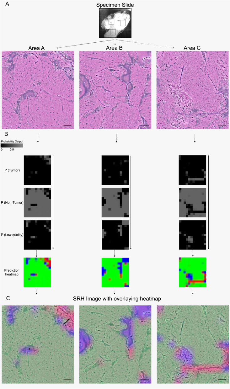
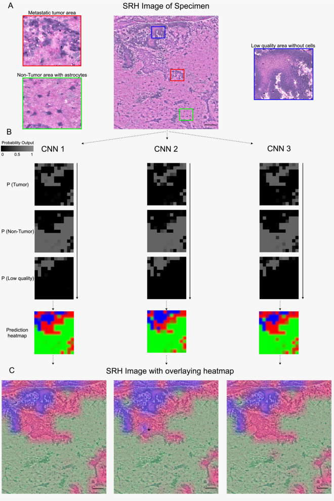
Determining the presence of tumor in biopsies and the decision-making during resections is often dependent on intraoperative rapid frozen-section histopathology. Recently, stimulated Raman scattering microscopy has been introduced to rapidly generate digital hematoxylin-and-eosin-stained-like images (stimulated Raman histology) for intraoperative analysis. To enable intraoperative prediction of tumor presence, we aimed to develop a new deep residual convolutional neural network in an automated pipeline and tested its validity. In a monocentric prospective clinical study with 94 patients undergoing biopsy, brain or spinal tumor resection, Stimulated Raman histology images of intraoperative tissue samples were obtained using a fiber-laser-based stimulated Raman scattering microscope. A residual network was established and trained in ResNetV50 to predict three classes for each image: (1) tumor, (2) non-tumor, and (3) low-quality. The residual network was validated on images obtained in three small random areas within the tissue samples and were blindly independently reviewed by a neuropathologist as ground truth. 402 images derived from 132 tissue samples were analyzed representing the entire spectrum of neurooncological surgery. The automated workflow took in a mean of 240 s per case, and the residual network correctly classified tumor (305/326), non-tumorous tissue (49/67), and low-quality (6/9) images with an inter-rater agreement of 89.6% (κ = 0.671). An excellent internal consistency was found among the random areas with 90.2% (Cα = 0.942) accuracy. In conclusion, the novel stimulated Raman histology-based residual network can reliably detect the microscopic presence of tumor and differentiate from non-tumorous brain tissue in resection and biopsy samples within 4 min and may pave a promising way for an alternative rapid intraoperative histopathological decision-making tool.

摘要

在活检中确定肿瘤的存在以及在切除过程中的决策通常依赖于术中快速冷冻切片组织病理学检查。最近,受激拉曼散射显微镜已被引入,以快速生成用于术中分析的数字苏木精-伊红染色样图像(受激拉曼组织学)。为了能够在术中预测肿瘤的存在,我们旨在开发一个新的深度残差卷积神经网络,并在自动流水线中进行测试。在一项 94 例接受活检、脑或脊髓肿瘤切除的患者的单中心前瞻性临床研究中,使用基于光纤激光的受激拉曼散射显微镜获取术中组织样本的受激拉曼组织学图像。建立了一个残差网络,并在 ResNetV50 中进行训练,以对每张图像预测三个类别:(1)肿瘤,(2)非肿瘤,和(3)低质量。该残差网络在组织样本内三个小随机区域获得的图像上进行验证,并由神经病理学家作为金标准进行盲法独立审查。共分析了来自 132 个组织样本的 402 张图像,代表了神经肿瘤学手术的整个范围。自动化工作流程平均每例耗时 240 秒,该残差网络正确分类了肿瘤(305/326)、非肿瘤组织(49/67)和低质量(6/9)图像,观察者间的一致性为 89.6%(κ=0.671)。在随机区域内发现了极好的内部一致性,准确率为 90.2%(Cα=0.942)。总之,新型基于受激拉曼组织学的残差网络可以可靠地检测肿瘤的微观存在,并在 4 分钟内从切除和活检样本中的非肿瘤脑组织中区分开来,可能为替代快速术中组织病理学决策工具铺平道路。

https://cdn.ncbi.nlm.nih.gov/pmc/blobs/1261/9356422/c88525695ef4/40478_2022_1411_Fig5_HTML.jpg
https://cdn.ncbi.nlm.nih.gov/pmc/blobs/1261/9356422/2109a8817873/40478_2022_1411_Fig1_HTML.jpg
https://cdn.ncbi.nlm.nih.gov/pmc/blobs/1261/9356422/91528d73f285/40478_2022_1411_Fig2_HTML.jpg
https://cdn.ncbi.nlm.nih.gov/pmc/blobs/1261/9356422/5e94a3b9c8a9/40478_2022_1411_Fig3_HTML.jpg
https://cdn.ncbi.nlm.nih.gov/pmc/blobs/1261/9356422/220c34c5dc39/40478_2022_1411_Fig4_HTML.jpg
https://cdn.ncbi.nlm.nih.gov/pmc/blobs/1261/9356422/c88525695ef4/40478_2022_1411_Fig5_HTML.jpg

相似文献

1
Novel rapid intraoperative qualitative tumor detection by a residual convolutional neural network using label-free stimulated Raman scattering microscopy.基于无标记受激拉曼散射显微镜的残余卷积神经网络实现术中快速定性肿瘤检测。
Acta Neuropathol Commun. 2022 Aug 6;10(1):109. doi: 10.1186/s40478-022-01411-x.
2
Streamlined Intraoperative Brain Tumor Classification and Molecular Subtyping in Stereotactic Biopsies Using Stimulated Raman Histology and Deep Learning.基于受激拉曼组织学和深度学习的立体定向活检术中简化脑肿瘤分类和分子亚型分析。
Clin Cancer Res. 2024 Sep 3;30(17):3824-3836. doi: 10.1158/1078-0432.CCR-23-3842.
3
Rapid histology of laryngeal squamous cell carcinoma with deep-learning based stimulated Raman scattering microscopy.基于深度学习的受激拉曼散射显微镜快速检测喉鳞状细胞癌。
Theranostics. 2019 Apr 13;9(9):2541-2554. doi: 10.7150/thno.32655. eCollection 2019.
4
Label-free brain tumor imaging using Raman-based methods.基于拉曼光谱的无标记脑肿瘤成像。
J Neurooncol. 2021 Feb;151(3):393-402. doi: 10.1007/s11060-019-03380-z. Epub 2021 Feb 21.
5
Neuropathological interpretation of stimulated Raman histology images of brain and spine tumors: part B.脑和脊柱肿瘤激发拉曼组织学图像的神经病理学解读:B 部分。
Neurosurg Rev. 2022 Apr;45(2):1721-1729. doi: 10.1007/s10143-021-01711-1. Epub 2021 Dec 10.
6
Rapid, label-free detection of diffuse glioma recurrence using intraoperative stimulated Raman histology and deep neural networks.利用术中受激拉曼组织学和深度神经网络快速、无标记检测弥漫性胶质瘤复发。
Neuro Oncol. 2021 Jan 30;23(1):144-155. doi: 10.1093/neuonc/noaa162.
7
Fast Real-Time Brain Tumor Detection Based on Stimulated Raman Histology and Self-Supervised Deep Learning Model.基于受激拉曼组织学和自监督深度学习模型的快速实时脑肿瘤检测。
J Imaging Inform Med. 2024 Jun;37(3):1160-1176. doi: 10.1007/s10278-024-01001-4. Epub 2024 Feb 7.
8
Stimulated Raman Scattering Microscopy Enables Gleason Scoring of Prostate Core Needle Biopsy by a Convolutional Neural Network.受激拉曼散射显微镜通过卷积神经网络实现前列腺核心针活检的 Gleason 评分。
Cancer Res. 2023 Feb 15;83(4):641-651. doi: 10.1158/0008-5472.CAN-22-2146.
9
Near real-time intraoperative brain tumor diagnosis using stimulated Raman histology and deep neural networks.利用受激拉曼组织学和深度神经网络进行近实时术中脑瘤诊断。
Nat Med. 2020 Jan;26(1):52-58. doi: 10.1038/s41591-019-0715-9. Epub 2020 Jan 6.
10
Advancements in Neurosurgical Intraoperative Histology.神经外科术中组织学进展。
Tomography. 2024 May 9;10(5):693-704. doi: 10.3390/tomography10050054.

引用本文的文献

1
Deep learning in neurosurgery: a systematic literature review with a structured analysis of applications across subspecialties.神经外科中的深度学习:一项系统的文献综述,并对各亚专业的应用进行结构化分析。
Front Neurol. 2025 Apr 16;16:1532398. doi: 10.3389/fneur.2025.1532398. eCollection 2025.
2
Confocal Laser Endomicroscopy: Enhancing Intraoperative Decision Making in Neurosurgery.共聚焦激光内镜显微镜:增强神经外科手术中的术中决策
Diagnostics (Basel). 2025 Feb 19;15(4):499. doi: 10.3390/diagnostics15040499.
3
Image Quality Assessment and Reliability Analysis of Artificial Intelligence-Based Tumor Classification of Stimulated Raman Histology of Tumor Biobank Samples.

本文引用的文献

1
Rapid Automated Analysis of Skull Base Tumor Specimens Using Intraoperative Optical Imaging and Artificial Intelligence.术中光学成像和人工智能快速分析颅底肿瘤标本
Neurosurgery. 2022 Jun 1;90(6):758-767. doi: 10.1227/neu.0000000000001929. Epub 2022 Mar 30.
2
Confocal Laser Microscopy in Neurosurgery: State of the Art of Actual Clinical Applications.神经外科中的共聚焦激光显微镜:实际临床应用的现状
J Clin Med. 2021 May 10;10(9):2035. doi: 10.3390/jcm10092035.
3
Frozen Section Quality Assurance.冰冻切片质量保证
肿瘤生物样本库样本的受激拉曼组织学人工智能辅助肿瘤分类的图像质量评估与可靠性分析
Diagnostics (Basel). 2024 Nov 30;14(23):2701. doi: 10.3390/diagnostics14232701.
4
Artificial Intelligence-Assisted Stimulated Raman Histology: New Frontiers in Vibrational Tissue Imaging.人工智能辅助受激拉曼组织学:振动组织成像的新前沿
Cancers (Basel). 2024 Nov 22;16(23):3917. doi: 10.3390/cancers16233917.
5
Fast intraoperative detection of primary central nervous system lymphoma and differentiation from common central nervous system tumors using stimulated Raman histology and deep learning.使用受激拉曼组织学和深度学习在术中快速检测原发性中枢神经系统淋巴瘤并与常见中枢神经系统肿瘤进行鉴别。
Neuro Oncol. 2025 Jun 21;27(5):1297-1310. doi: 10.1093/neuonc/noae270.
6
Fast intraoperative detection of primary CNS lymphoma and differentiation from common CNS tumors using stimulated Raman histology and deep learning.使用受激拉曼组织学和深度学习在术中快速检测原发性中枢神经系统淋巴瘤并与常见中枢神经系统肿瘤进行鉴别。
medRxiv. 2024 Aug 26:2024.08.25.24312509. doi: 10.1101/2024.08.25.24312509.
7
Practical Application of Deep Learning in Diagnostic Neuropathology-Reimagining a Histological Asset in the Era of Precision Medicine.深度学习在诊断神经病理学中的实际应用——在精准医学时代重塑一种组织学资源
Cancers (Basel). 2024 May 23;16(11):1976. doi: 10.3390/cancers16111976.
8
[Intraoperative stimulated Raman histology for personalized brain tumor surgery].[术中受激拉曼组织学用于个性化脑肿瘤手术]
Chirurgie (Heidelb). 2024 Apr;95(4):274-279. doi: 10.1007/s00104-024-02038-5. Epub 2024 Feb 9.
9
Machine Learning in Neurosurgery: Toward Complex Inputs, Actionable Predictions, and Generalizable Translations.神经外科中的机器学习:迈向复杂输入、可操作预测和可推广转化
Cureus. 2024 Jan 9;16(1):e51963. doi: 10.7759/cureus.51963. eCollection 2024 Jan.
10
Virtual Staining of Nonfixed Tissue Histology.非固定组织学的虚拟染色。
Mod Pathol. 2024 May;37(5):100444. doi: 10.1016/j.modpat.2024.100444. Epub 2024 Feb 6.
Am J Clin Pathol. 2021 Aug 4;156(3):461-470. doi: 10.1093/ajcp/aqaa259.
4
Brain Shift in Neuronavigation of Brain Tumors: An Updated Review of Intra-Operative Ultrasound Applications.脑肿瘤神经导航中的脑移位:术中超声应用的最新综述
Front Oncol. 2021 Feb 8;10:618837. doi: 10.3389/fonc.2020.618837. eCollection 2020.
5
Intraoperative MRI for Brain Tumors.脑肿瘤术中磁共振成像。
J Neurooncol. 2021 Feb;151(3):479-490. doi: 10.1007/s11060-020-03667-6. Epub 2021 Feb 21.
6
Label-free brain tumor imaging using Raman-based methods.基于拉曼光谱的无标记脑肿瘤成像。
J Neurooncol. 2021 Feb;151(3):393-402. doi: 10.1007/s11060-019-03380-z. Epub 2021 Feb 21.
7
Intraoperative Confocal Laser Endomicroscopy Examination of Tissue Microstructure During Fluorescence-Guided Brain Tumor Surgery.荧光引导下脑肿瘤手术中组织微观结构的术中共聚焦激光内镜检查
Front Oncol. 2020 Dec 4;10:599250. doi: 10.3389/fonc.2020.599250. eCollection 2020.
8
Intraoperative Imaging for High-Grade Glioma Surgery.术中影像学在高级别胶质瘤手术中的应用。
Neurosurg Clin N Am. 2021 Jan;32(1):47-54. doi: 10.1016/j.nec.2020.09.003. Epub 2020 Nov 5.
9
Rapid, label-free detection of diffuse glioma recurrence using intraoperative stimulated Raman histology and deep neural networks.利用术中受激拉曼组织学和深度神经网络快速、无标记检测弥漫性胶质瘤复发。
Neuro Oncol. 2021 Jan 30;23(1):144-155. doi: 10.1093/neuonc/noaa162.
10
An automated tissue-to-diagnosis pipeline using intraoperative stimulated Raman histology and deep learning.一种使用术中受激拉曼组织学和深度学习的自动化组织诊断流程。
Mol Cell Oncol. 2020 Apr 1;7(3):1736742. doi: 10.1080/23723556.2020.1736742. eCollection 2020.