Center for Neurodegeneration and Experimental Therapeutics, Department of Neurology, The University of Alabama at Birmingham (UAB), 1719 6th Ave. South, CIRC 446, Birmingham, AL, 35294-0021, USA.
Department of Neurological Sciences, Rush University Medical Center, Chicago, IL, 60612, USA.
Acta Neuropathol. 2020 May;139(5):855-874. doi: 10.1007/s00401-020-02126-w. Epub 2020 Jan 29.
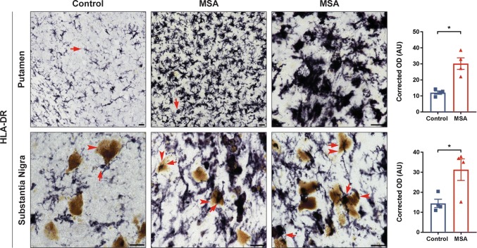
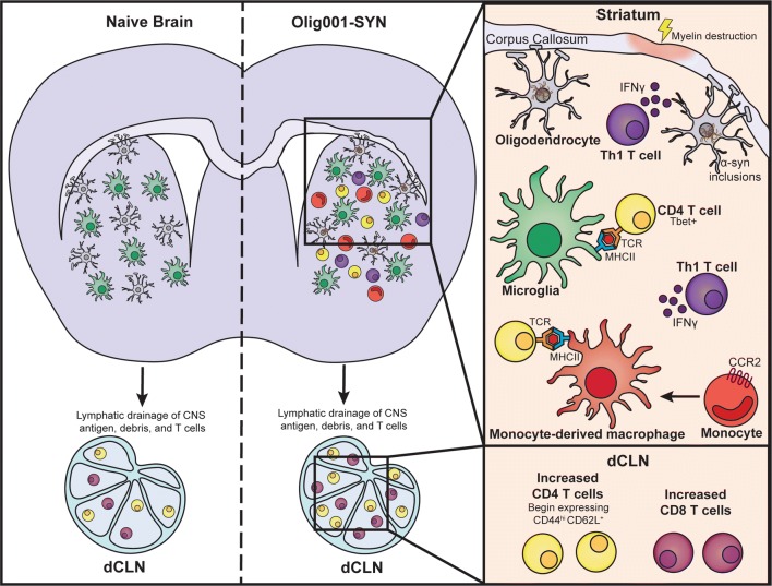
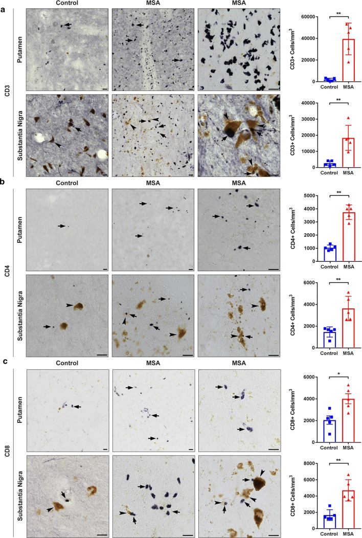
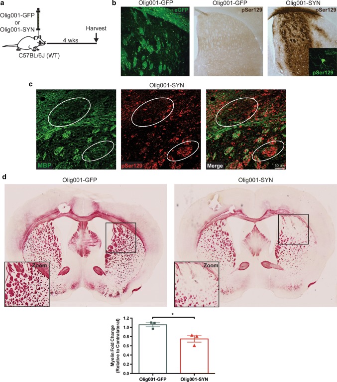
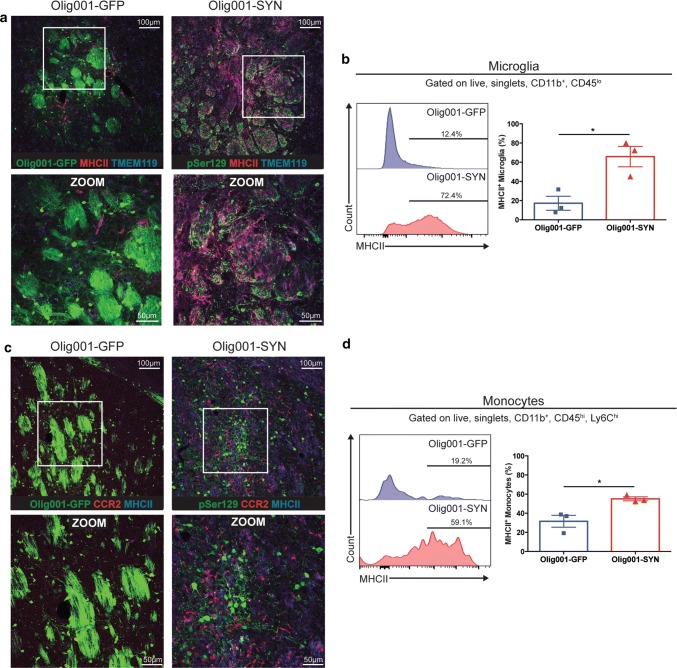
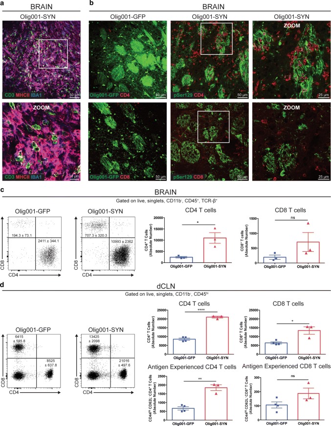
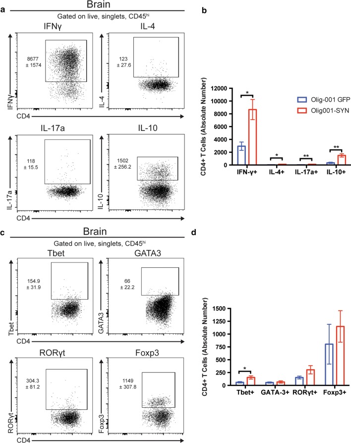
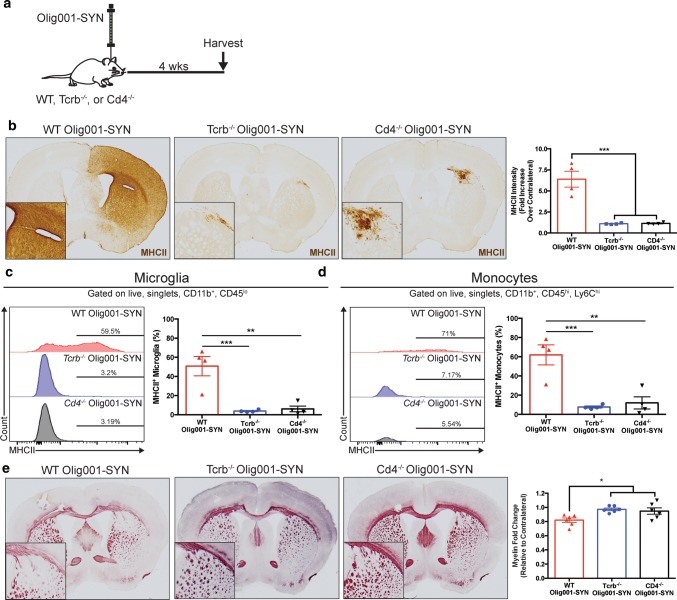
Multiple system atrophy (MSA) is a progressive neurodegenerative disorder characterized by abnormal accumulation of alpha-synuclein (α-syn) in oligodendrocytes accompanied by inflammation, demyelination, and subsequent synapse and neuronal loss. Little is known about the mechanisms of neurodegeneration in MSA. However, recent work has highlighted the important role of the immune system to the pathophysiology of other synuclein-related diseases such as Parkinson's disease. In this study, we investigated postmortem brain tissue from MSA patients and control subjects for evidence of immune activation in the brain. We found a significant increase of HLA-DR microglia in the putamen and substantia nigra of MSA patient tissue compared to controls, as well as significant increases in CD3, CD4, and CD8 T cells in these same brain regions. To model MSA in vivo, we utilized a viral vector that selectively overexpresses α-syn in oligodendrocytes (Olig001-SYN) with > 95% tropism in the dorsal striatum of mice, resulting in demyelination and neuroinflammation similar to that observed in human MSA. Oligodendrocyte transduction with this vector resulted in a robust inflammatory response, which included increased MHCII expression on central nervous system (CNS) resident microglia, and infiltration of pro-inflammatory monocytes into the CNS. We also observed robust infiltration of CD4 T cells into the CNS and antigen-experienced CD4 T cells in the draining cervical lymph nodes. Importantly, genetic deletion of TCR-β or CD4 T cells attenuated α-syn-induced inflammation and demyelination in vivo. These results suggest that T cell priming and infiltration into the CNS are key mechanisms of disease pathogenesis in MSA, and therapeutics targeting T cells may be disease modifying.
多系统萎缩症(MSA)是一种进行性神经退行性疾病,其特征是少突胶质细胞中异常积聚的α-突触核蛋白(α-syn),伴有炎症、脱髓鞘以及随后的突触和神经元丢失。目前对于 MSA 中神经退行性变的机制知之甚少。然而,最近的研究强调了免疫系统对其他与突触核蛋白相关疾病(如帕金森病)的病理生理学的重要作用。在这项研究中,我们研究了 MSA 患者和对照受试者的死后脑组织,以寻找大脑中免疫激活的证据。我们发现,与对照组相比,MSA 患者组织纹状体和黑质中的 HLA-DR 小胶质细胞显著增加,并且这些相同脑区中的 CD3、CD4 和 CD8 T 细胞也显著增加。为了在体内模拟 MSA,我们利用一种病毒载体,该载体选择性地在少突胶质细胞中过表达 α-syn(Olig001-SYN),在小鼠背侧纹状体中的靶向性超过 95%,导致脱髓鞘和神经炎症,类似于人类 MSA 中观察到的。该载体对少突胶质细胞的转导导致强烈的炎症反应,包括中枢神经系统(CNS)固有小胶质细胞上 MHCII 表达的增加,以及促炎单核细胞向 CNS 的浸润。我们还观察到 CD4 T 细胞大量浸润 CNS 和引流颈淋巴结中的抗原经验 CD4 T 细胞。重要的是,TCR-β或 CD4 T 细胞的遗传缺失减轻了体内α-syn 诱导的炎症和脱髓鞘。这些结果表明,T 细胞的初始激活和浸润到中枢神经系统是 MSA 发病机制的关键机制,针对 T 细胞的治疗方法可能具有疾病修饰作用。