Department of Radiology, Charité-Universitätsmedizin Berlin, Germany.
Neuroimage Clin. 2012 Sep 12;1(1):81-90. doi: 10.1016/j.nicl.2012.09.003. eCollection 2012.
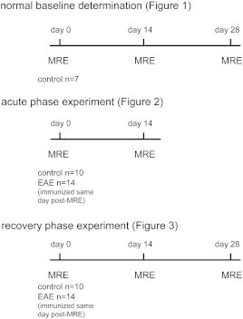
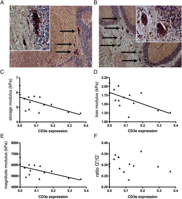
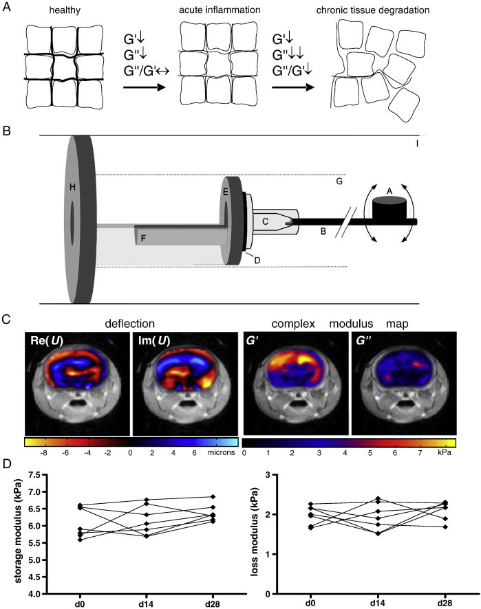
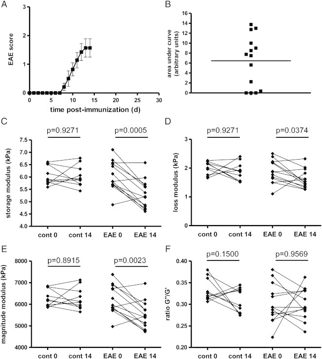
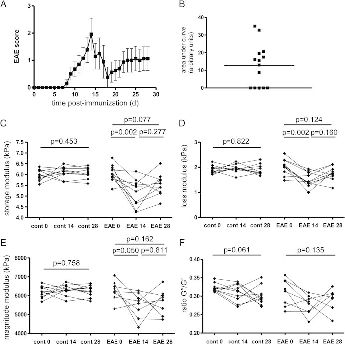
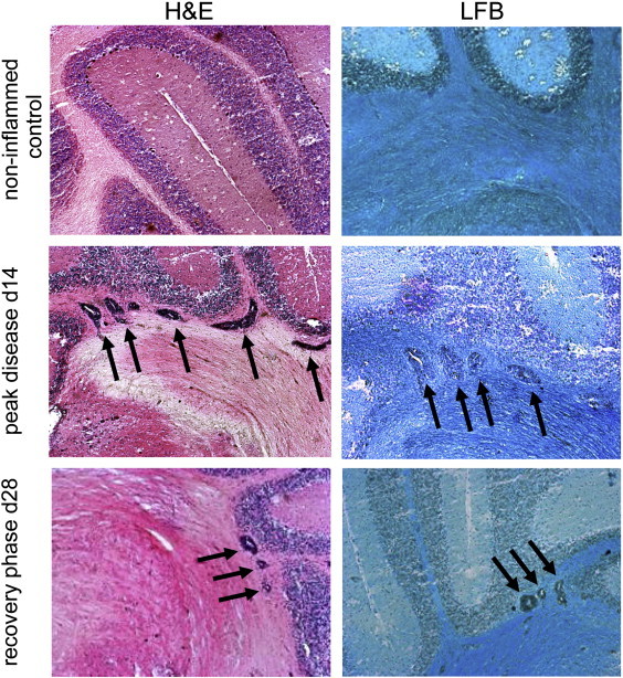
Cerebral magnetic resonance elastography (MRE) measures the viscoelastic properties of brain tissues in vivo. It was recently shown that brain viscoelasticity is reduced in patients with multiple sclerosis (MS), highlighting the potential of cerebral MRE to detect tissue pathology during neuroinflammation. To further investigate the relationship between inflammation and brain viscoelasticity, we applied MRE to a mouse model of MS, experimental autoimmune encephalomyelitis (EAE). EAE was induced and monitored by MRE in a 7-tesla animal MRI scanner over 4 weeks. At the peak of the disease (day 14 after immunization), we detected a significant decrease in both the storage modulus (G') and the loss modulus (G″), indicating that both the elasticity and the viscosity of the brain are reduced during acute inflammation. Interestingly, these parameters normalized at a later time point (day 28) corresponding to the clinical recovery phase. Consistent with this, we observed a clear correlation between viscoelastic tissue alteration and the magnitude of perivascular T cell infiltration at both day 14 and day 28. Hence, acute neuroinflammation is associated with reduced mechanical cohesion of brain tissues. Moreover, the reduction of brain viscoelasticity appears to be a reversible process, which is restored when inflammation resolves. For the first time, our study has demonstrated the applicability of cerebral MRE in EAE, and showed that this novel imaging technology is highly sensitive to early tissue alterations resulting from the inflammatory processes. Thus, MRE may serve to monitor early stages of perivascular immune infiltration during neuroinflammation.
脑磁共振弹性成像(MRE)测量脑组织的黏弹性。最近的研究表明,多发性硬化症(MS)患者的脑黏弹性降低,突出了 MRE 检测神经炎症期间组织病理学的潜力。为了进一步研究炎症与脑黏弹性之间的关系,我们将 MRE 应用于实验性自身免疫性脑脊髓炎(EAE)的 MS 小鼠模型。EAE 在 7T 动物 MRI 扫描仪中通过 MRE 诱导和监测,为期 4 周。在疾病高峰期(免疫后第 14 天),我们检测到存储模量(G')和损耗模量(G")均显著降低,表明急性炎症期间大脑的弹性和粘性均降低。有趣的是,这些参数在稍后的时间点(免疫后第 28 天)即临床恢复期正常化。与此一致,我们在第 14 天和第 28 天观察到黏弹性组织改变与血管周围 T 细胞浸润程度之间存在明显的相关性。因此,急性神经炎症与脑组织机械结合力降低有关。此外,脑黏弹性降低似乎是一个可逆的过程,当炎症消退时会恢复。本研究首次证明了脑 MRE 在 EAE 中的适用性,并表明这种新型成像技术对炎症过程引起的早期组织改变非常敏感。因此,MRE 可能用于监测神经炎症期间血管周围免疫浸润的早期阶段。