Institute for Physical Science and Technology, University of Maryland, College Park, MD 20742.
University of Maryland School of Medicine, Baltimore, MD 21201.
Mol Biol Cell. 2020 Jul 21;31(16):1753-1764. doi: 10.1091/mbc.E19-11-0614. Epub 2020 Feb 5.
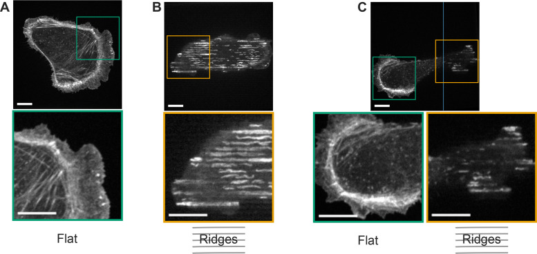
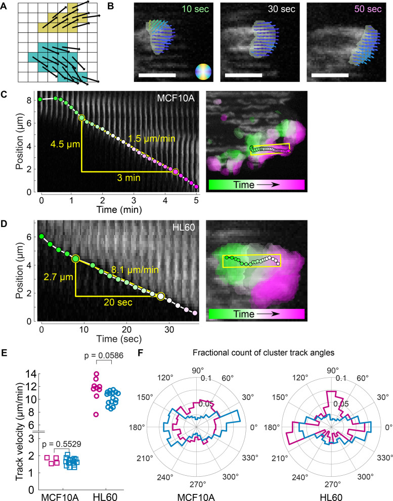
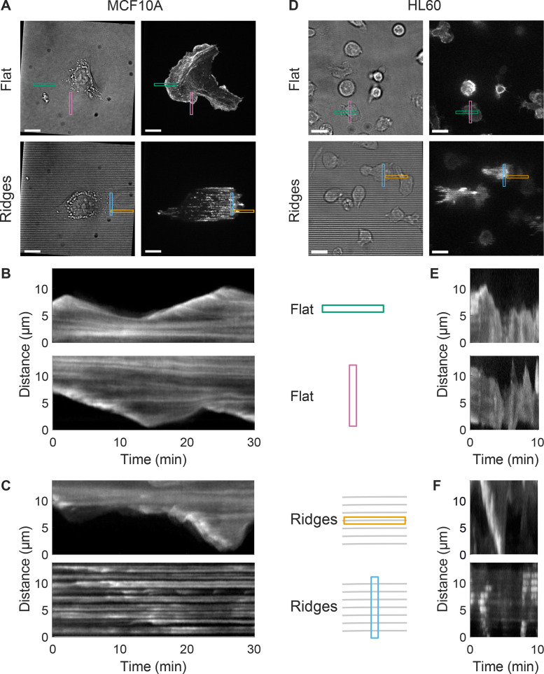
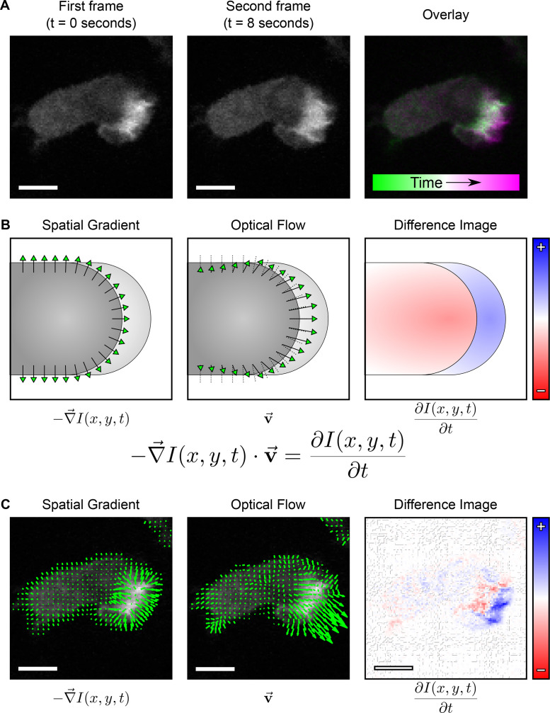
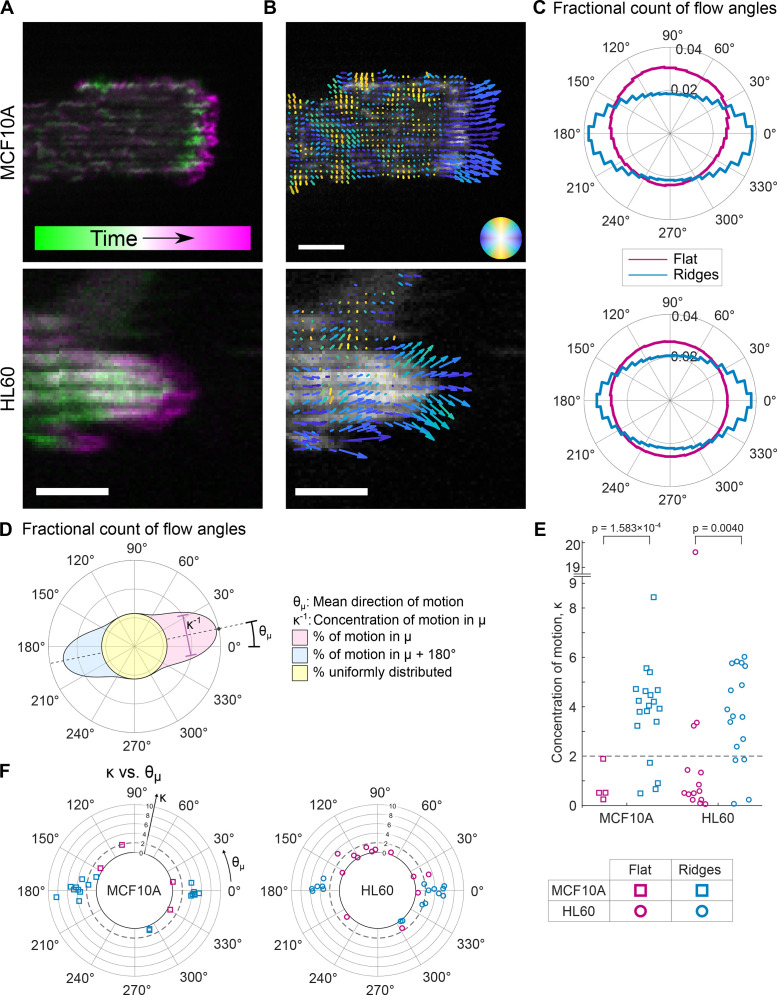
The dynamic rearrangement of the actin cytoskeleton is an essential component of many mechanotransduction and cellular force generation pathways. Here we use periodic surface topographies with feature sizes comparable to those of in vivo collagen fibers to measure and compare actin dynamics for two representative cell types that have markedly different migratory modes and physiological purposes: slowly migrating epithelial MCF10A cells and polarizing, fast-migrating, neutrophil-like HL60 cells. Both cell types exhibit reproducible guidance of actin waves (esotaxis) on these topographies, enabling quantitative comparisons of actin dynamics. We adapt a computer-vision algorithm, optical flow, to measure the directions of actin waves at the submicron scale. Clustering the optical flow into regions that move in similar directions enables micron-scale measurements of actin-wave speed and direction. Although the speed and morphology of actin waves differ between MCF10A and HL60 cells, the underlying actin guidance by nanotopography is similar in both cell types at the micron and submicron scales.
细胞骨架的动态重排是许多机械转导和细胞力产生途径的重要组成部分。在这里,我们使用周期性的表面形貌,其特征尺寸与体内胶原纤维相当,来测量和比较两种具有明显不同迁移模式和生理功能的代表性细胞类型的肌动蛋白动力学:迁移缓慢的上皮 MCF10A 细胞和极化的、快速迁移的、类似中性粒细胞的 HL60 细胞。这两种细胞类型在这些形貌上都表现出可重复的肌动蛋白波的导向(趋边运动),从而能够对肌动蛋白动力学进行定量比较。我们采用计算机视觉算法——光流法,在亚微米尺度上测量肌动蛋白波的方向。将光流聚类成以相似方向运动的区域,能够实现肌动蛋白波速度和方向的微米级测量。尽管 MCF10A 和 HL60 细胞之间的肌动蛋白波的速度和形态不同,但在微米和亚微米尺度上,纳米形貌对肌动蛋白的导向在这两种细胞类型中是相似的。