• 文献检索
  • 文档翻译
  • 深度研究
  • 学术资讯
  • Suppr Zotero 插件Zotero 插件
  • 邀请有礼
  • 套餐&价格
  • 历史记录
应用&插件
Suppr Zotero 插件Zotero 插件浏览器插件Mac 客户端Windows 客户端微信小程序
定价
高级版会员购买积分包购买API积分包
服务
文献检索文档翻译深度研究API 文档MCP 服务
关于我们
关于 Suppr公司介绍联系我们用户协议隐私条款
关注我们

Suppr 超能文献

核心技术专利:CN118964589B侵权必究
粤ICP备2023148730 号-1Suppr @ 2026

文献检索

告别复杂PubMed语法,用中文像聊天一样搜索,搜遍4000万医学文献。AI智能推荐,让科研检索更轻松。

立即免费搜索

文件翻译

保留排版,准确专业,支持PDF/Word/PPT等文件格式,支持 12+语言互译。

免费翻译文档

深度研究

AI帮你快速写综述,25分钟生成高质量综述,智能提取关键信息,辅助科研写作。

立即免费体验

节段性定量单(压力)门控灌注 CMR 提高了检测阻塞性冠状动脉疾病的诊断准确性。

Sub-segmental quantification of single (stress)-pass perfusion CMR improves the diagnostic accuracy for detection of obstructive coronary artery disease.

机构信息

Institute for Experimental and Translational Cardiovascular Imaging, University Hospital Frankfurt, Theodor-Stern Kai 7, 60590, Frankfurt am Main, Germany.

Department of Biomedical Sciences and Morphological and Functional Imaging, G. Martino University Hospital Messina, Via Consolare Valeria 1, Messina, 98100, Italy.

出版信息

J Cardiovasc Magn Reson. 2020 Feb 6;22(1):14. doi: 10.1186/s12968-020-0600-1.

DOI:10.1186/s12968-020-0600-1
PMID:32028980
原文链接:https://pmc.ncbi.nlm.nih.gov/articles/PMC7006214/
Abstract

BACKGROUND

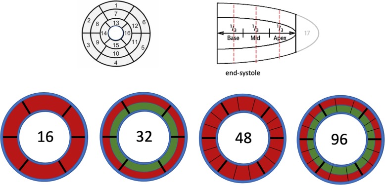
Myocardial perfusion with cardiovascular magnetic resonance (CMR) imaging is an established diagnostic test for evaluation of myocardial ischaemia. For quantification purposes, the 16 segment American Heart Association (AHA) model poses limitations in terms of extracting relevant information on the extent/severity of ischaemia as perfusion deficits will not always fall within an individual segment, which reduces its diagnostic value, and makes an accurate assessment of outcome data or a result comparison across various studies difficult. We hypothesised that division of the myocardial segments into epi- and endocardial layers and a further circumferential subdivision, resulting in a total of 96 segments, would improve the accuracy of detecting myocardial hypoperfusion. Higher (sub-)subsegmental recording of perfusion abnormalities, which are defined relatively to the normal reference using the subsegment with the highest value, may improve the spatial encoding of myocardial blood flow, based on a single stress perfusion acquisition.

OBJECTIVE

A proof of concept comparison study of subsegmentation approaches based on transmural segments (16 AHA and 48 segments) vs. subdivision into epi- and endocardial (32) subsegments vs. further circumferential subdivision into 96 (sub-)subsegments for diagnostic accuracy against invasively defined obstructive coronary artery disease (CAD).

METHODS

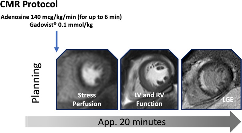
Thirty patients with obstructive CAD and 20 healthy controls underwent perfusion stress CMR imaging at 3 T during maximal adenosine vasodilation and a dual bolus injection of 0.1 mmol/kg gadobutrol. Using Fermi deconvolution for blood flow estimation, (sub-)subsegmental values were expressed relative to the (sub-)subsegment with the highest flow. In addition, endo-/epicardial flow ratios were calculated based on 32 and 96 (sub-)subsegments. A receiver operating characteristics (ROC) curve analysis was performed to compare the diagnostic performance of discrimination between patients with CAD and healthy controls. Observer reproducibility was assessed using Bland-Altman approaches.

RESULTS

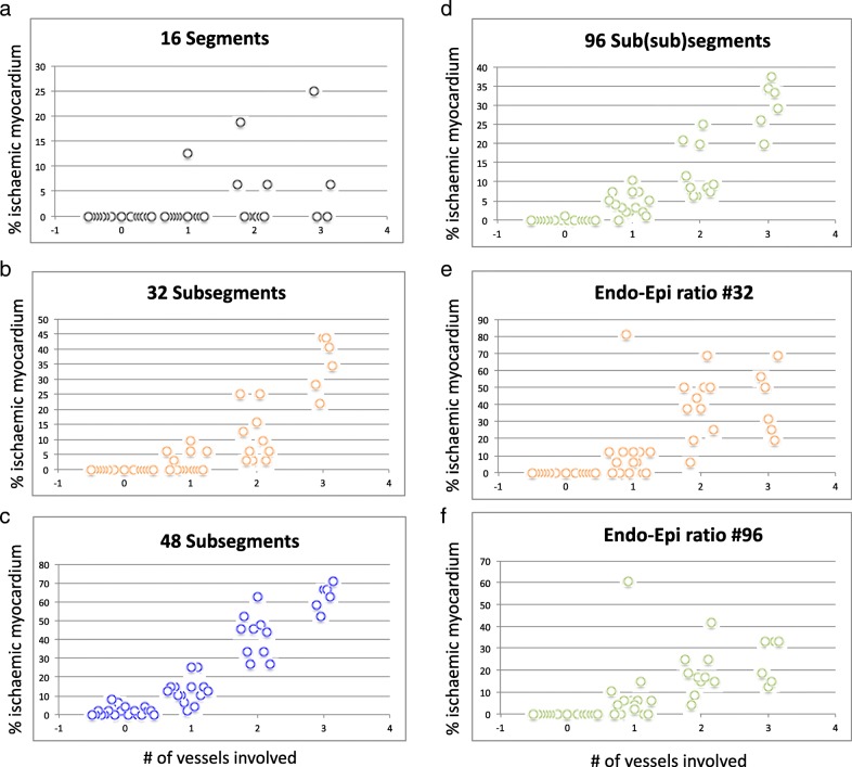
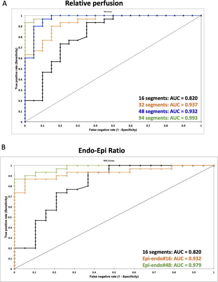
Subdivision into more and smaller segments revealed greater accuracy for #32, #48 and # 96 compared to the standard #16 approach (area under the curve (AUC): 0.937, 0.973 and 0.993 vs 0.820, p < 0.05). The #96-based endo-/epicardial ratio was superior to the #32 endo-/epicardial ratio (AUC 0.979, vs. 0.932, p < 0.05). Measurements for the #16 model showed marginally better reproducibility compared to #32, #48 and #96 (mean difference ± standard deviation: 2.0 ± 3.6 vs. 2.3 ± 4.0 vs 2.5 ± 4.4 vs. 4.1 ± 5.6).

CONCLUSIONS

Subsegmentation of the myocardium improves diagnostic accuracy and facilitates an objective cut-off-based description of hypoperfusion, and facilitates an objective description of hypoperfusion, including the extent and severity of myocardial ischaemia. Quantification based on a single (stress-only) pass reduces the overall amount of gadolinium contrast agent required and the length of the overall diagnostic study.

摘要

背景

心血管磁共振(CMR)心肌灌注成像已成为评估心肌缺血的既定诊断测试。出于定量目的,美国心脏协会(AHA)的 16 节段模型在提取关于缺血程度/严重程度的相关信息方面存在局限性,因为灌注缺损并不总是位于单个节段内,这降低了其诊断价值,并使得对结果数据进行准确评估或在不同研究之间进行结果比较变得困难。我们假设将心肌节段划分为心外膜和心内膜层,并进一步进行圆周细分,总共产生 96 个节段,将提高检测心肌低灌注的准确性。较高的(亚)节段灌注异常记录,相对于使用具有最高值的亚节段定义为相对正常参考值,可能会基于单次应激灌注采集提高心肌血流的空间编码。

目的

基于透壁节段(16 个 AHA 和 48 个节段)与心外膜和心内膜(32 个)细分与进一步圆周细分成 96 个(亚)节段(32 个)的亚节段方法的概念验证比较研究,以针对侵入性定义的阻塞性冠状动脉疾病(CAD)的诊断准确性。

方法

30 例阻塞性 CAD 患者和 20 例健康对照者在 3T 下接受最大腺苷血管扩张和 0.1mmol/kg 钆布醇双重团注的应激 CMR 成像。使用费米反卷积法进行血流估计,(亚)节段值相对于具有最高血流的(亚)节段表示。此外,还基于 32 和 96 个(亚)节段计算了心外膜/心内膜的血流比值。进行了接收器操作特征(ROC)曲线分析,以比较区分 CAD 患者和健康对照者的诊断性能。使用 Bland-Altman 方法评估观察者的可重复性。

结果

与标准的 16 节段方法相比,更多和更小的细分节段显示出更高的准确性(#32、#48 和#96 的曲线下面积(AUC)分别为 0.937、0.973 和 0.993 与 0.820,p < 0.05)。基于#96 的心外膜/心内膜比值优于基于#32 的心外膜/心内膜比值(AUC 0.979,vs. 0.932,p < 0.05)。与#32、#48 和#96 相比,#16 模型的测量值显示出略好的可重复性(平均差异 ± 标准差:2.0 ± 3.6 与 2.3 ± 4.0 与 2.5 ± 4.4 与 4.1 ± 5.6)。

结论

心肌细分提高了诊断准确性,并有助于客观地描述低灌注,包括心肌缺血的程度和严重程度。基于单次(仅应激)通过的定量减少了所需的总体钆造影剂量和整体诊断研究的长度。

https://cdn.ncbi.nlm.nih.gov/pmc/blobs/bb0d/7006214/7e6d325dd20e/12968_2020_600_Fig4_HTML.jpg
https://cdn.ncbi.nlm.nih.gov/pmc/blobs/bb0d/7006214/b8dcc29be7ad/12968_2020_600_Fig1_HTML.jpg
https://cdn.ncbi.nlm.nih.gov/pmc/blobs/bb0d/7006214/9cfb8ddfc625/12968_2020_600_Fig2_HTML.jpg
https://cdn.ncbi.nlm.nih.gov/pmc/blobs/bb0d/7006214/4aaa3677793d/12968_2020_600_Fig3_HTML.jpg
https://cdn.ncbi.nlm.nih.gov/pmc/blobs/bb0d/7006214/7e6d325dd20e/12968_2020_600_Fig4_HTML.jpg

相似文献

1
Sub-segmental quantification of single (stress)-pass perfusion CMR improves the diagnostic accuracy for detection of obstructive coronary artery disease.节段性定量单(压力)门控灌注 CMR 提高了检测阻塞性冠状动脉疾病的诊断准确性。
J Cardiovasc Magn Reson. 2020 Feb 6;22(1):14. doi: 10.1186/s12968-020-0600-1.
2
Quantitative assessment of myocardial blood flow in coronary artery disease by cardiovascular magnetic resonance: comparison of Fermi and distributed parameter modeling against invasive methods.通过心血管磁共振对冠状动脉疾病心肌血流进行定量评估:费米模型和分布参数模型与侵入性方法的比较
J Cardiovasc Magn Reson. 2016 Sep 13;18(1):57. doi: 10.1186/s12968-016-0270-1.
3
Utility of stress perfusion-cardiac magnetic resonance in follow-up of patients undergoing percutaneous coronary interventions of the left main coronary artery.应激灌注心脏磁共振成像在左主干冠状动脉经皮冠状动脉介入治疗患者随访中的应用价值
Int J Cardiovasc Imaging. 2017 Oct;33(10):1589-1597. doi: 10.1007/s10554-017-1149-4. Epub 2017 Apr 28.
4
A comparison of cardiovascular magnetic resonance and single photon emission computed tomography (SPECT) perfusion imaging in left main stem or equivalent coronary artery disease: a CE-MARC substudy.左主干或等效冠状动脉疾病中心血管磁共振与单光子发射计算机断层扫描(SPECT)灌注成像的比较:CE-MARC 子研究。
J Cardiovasc Magn Reson. 2017 Nov 6;19(1):84. doi: 10.1186/s12968-017-0398-7.
5
Adenosine stress CMR T1-mapping detects early microvascular dysfunction in patients with type 2 diabetes mellitus without obstructive coronary artery disease.腺苷负荷心脏磁共振 T1 mapping 检测 2 型糖尿病无阻塞性冠状动脉疾病患者的早期微血管功能障碍。
J Cardiovasc Magn Reson. 2017 Oct 25;19(1):81. doi: 10.1186/s12968-017-0397-8.
6
Combined Assessment of Stress Myocardial Perfusion Cardiovascular Magnetic Resonance and Flow Measurement in the Coronary Sinus Improves Prediction of Functionally Significant Coronary Stenosis Determined by Fractional Flow Reserve in Multivessel Disease.联合评估应激心肌灌注心血管磁共振和冠状窦血流测量可提高多血管病变中通过血流储备分数测定的功能性显著冠状动脉狭窄的预测。
J Am Heart Assoc. 2018 Jan 26;7(3):e007736. doi: 10.1161/JAHA.117.007736.
7
Diagnostic value of global myocardial perfusion reserve assessment based on coronary sinus flow measurements using cardiovascular magnetic resonance in addition to myocardial stress perfusion imaging.基于冠状动脉窦血流测量的心肌整体灌注储备评估在心肌负荷灌注成像之外的诊断价值。
Eur Heart J Cardiovasc Imaging. 2017 May 1;18(8):851-859. doi: 10.1093/ehjci/jew315.
8
Automated Pixel-Wise Quantitative Myocardial Perfusion Mapping by CMR to Detect Obstructive Coronary Artery Disease and Coronary Microvascular Dysfunction: Validation Against Invasive Coronary Physiology.CMR 自动像素定量心肌灌注成像检测冠状动脉疾病和冠状动脉微血管功能障碍:与有创性冠状动脉生理学的对照验证。
JACC Cardiovasc Imaging. 2019 Oct;12(10):1958-1969. doi: 10.1016/j.jcmg.2018.12.022. Epub 2019 Feb 13.
9
Measurement of myocardial blood flow by cardiovascular magnetic resonance perfusion: comparison of distributed parameter and Fermi models with single and dual bolus.心血管磁共振灌注测量心肌血流:分布参数模型和费米模型在单团注与双团注情况下的比较
J Cardiovasc Magn Reson. 2015 Feb 17;17(1):17. doi: 10.1186/s12968-015-0125-1.
10
Fully quantitative pixel-wise analysis of cardiovascular magnetic resonance perfusion improves discrimination of dark rim artifact from perfusion defects associated with epicardial coronary stenosis.对心血管磁共振灌注的完全定量像素分析可改善对与心外膜冠状动脉狭窄相关的暗带伪影与灌注缺损的区分。
J Cardiovasc Magn Reson. 2018 Mar 8;20(1):16. doi: 10.1186/s12968-018-0436-0.

引用本文的文献

1
Correlation between cardiac and hepatic native T1 value and myocardial late gadolinium enhancement in children with Kawasaki disease.川崎病患儿心脏与肝脏固有T1值及心肌延迟钆增强之间的相关性
Quant Imaging Med Surg. 2025 Mar 3;15(3):1990-2002. doi: 10.21037/qims-24-791. Epub 2025 Feb 26.
2
Using artificial intelligence and promoter-level transcriptome analysis to identify a biomarker as a possible prognostic predictor of cardiac complications in male patients with Fabry disease.利用人工智能和启动子水平的转录组分析来鉴定一种生物标志物,作为男性法布里病患者心脏并发症可能的预后预测指标。
Mol Genet Metab Rep. 2024 Oct 13;41:101152. doi: 10.1016/j.ymgmr.2024.101152. eCollection 2024 Dec.
3

本文引用的文献

1
2019 ESC Guidelines for the diagnosis and management of chronic coronary syndromes.2019年欧洲心脏病学会慢性冠状动脉综合征诊断和管理指南
Eur Heart J. 2020 Jan 14;41(3):407-477. doi: 10.1093/eurheartj/ehz425.
2
Controversies in Diagnostic Imaging of Patients With Suspected Stable and Acute Chest Pain Syndromes.疑似稳定型和急性胸痛综合征患者的诊断影像学争议。
JACC Cardiovasc Imaging. 2019 Jul;12(7 Pt 1):1254-1278. doi: 10.1016/j.jcmg.2019.05.009.
3
Magnetic Resonance Perfusion or Fractional Flow Reserve in Coronary Disease.磁共振灌注或冠状动脉疾病的血流储备分数。
High-resolution quantification of stress perfusion defects by cardiac magnetic resonance.
通过心脏磁共振对压力灌注缺损进行高分辨率定量分析。
Eur Heart J Imaging Methods Pract. 2024 Jan 9;2(1):qyae001. doi: 10.1093/ehjimp/qyae001. eCollection 2024 Jan.
4
Automatic calculation of myocardial perfusion reserve using deep learning with uncertainty quantification.使用具有不确定性量化的深度学习自动计算心肌灌注储备。
Quant Imaging Med Surg. 2023 Dec 1;13(12):7936-7949. doi: 10.21037/qims-23-840. Epub 2023 Oct 10.
5
MR imaging of primary benign cardiac tumors in the pediatric population.儿科原发性良性心脏肿瘤的磁共振成像
Heliyon. 2023 Sep 12;9(9):e19932. doi: 10.1016/j.heliyon.2023.e19932. eCollection 2023 Sep.
6
2021 - State of our JCMR.2021年——我们的《日本临床肿瘤学杂志》现状
J Cardiovasc Magn Reson. 2022 Mar 4;24(1):14. doi: 10.1186/s12968-021-00840-2.
7
Multimodality Imaging in Ischemic Chronic Cardiomyopathy.缺血性慢性心肌病的多模态成像
J Imaging. 2022 Feb 1;8(2):35. doi: 10.3390/jimaging8020035.
8
Stress CMR in Known or Suspected CAD: Diagnostic and Prognostic Role.已知或疑似 CAD 患者的压力心脏磁共振:诊断和预后作用。
Biomed Res Int. 2021 Jan 14;2021:6678029. doi: 10.1155/2021/6678029. eCollection 2021.
9
Contemporary Cardiac MRI in Chronic Coronary Artery Disease.慢性冠状动脉疾病的当代心脏磁共振成像
Eur Cardiol. 2020 Jun 15;15:e50. doi: 10.15420/ecr.2019.17. eCollection 2020 Feb.
N Engl J Med. 2019 Jun 20;380(25):2418-2428. doi: 10.1056/NEJMoa1716734.
4
Society for Cardiovascular Magnetic Resonance (SCMR) expert consensus for CMR imaging endpoints in clinical research: part I - analytical validation and clinical qualification.心血管磁共振学会 (SCMR) 临床研究中 CMRI 终点的专家共识:第一部分——分析验证和临床资格认证。
J Cardiovasc Magn Reson. 2018 Sep 20;20(1):67. doi: 10.1186/s12968-018-0484-5.
5
2018 ESC/EACTS Guidelines on myocardial revascularization.2018年欧洲心脏病学会/欧洲心胸外科学会心肌血运重建指南。
Eur Heart J. 2019 Jan 7;40(2):87-165. doi: 10.1093/eurheartj/ehy394.
6
Fully quantitative pixel-wise analysis of cardiovascular magnetic resonance perfusion improves discrimination of dark rim artifact from perfusion defects associated with epicardial coronary stenosis.对心血管磁共振灌注的完全定量像素分析可改善对与心外膜冠状动脉狭窄相关的暗带伪影与灌注缺损的区分。
J Cardiovasc Magn Reson. 2018 Mar 8;20(1):16. doi: 10.1186/s12968-018-0436-0.
7
Diagnostic Performance of Fully Automated Pixel-Wise Quantitative Myocardial Perfusion Imaging by Cardiovascular Magnetic Resonance.心血管磁共振全自动像素定量心肌灌注成像的诊断性能。
JACC Cardiovasc Imaging. 2018 May;11(5):697-707. doi: 10.1016/j.jcmg.2018.01.005. Epub 2018 Feb 14.
8
Native T1 and ECV of Noninfarcted Myocardium and Outcome in Patients With Coronary Artery Disease.非梗死心肌的固有 T1 和 ECV 与冠状动脉疾病患者的结局。
J Am Coll Cardiol. 2018 Feb 20;71(7):766-778. doi: 10.1016/j.jacc.2017.12.020.
9
Definition of Left Ventricular Segments for Cardiac Magnetic Resonance Imaging.用于心脏磁共振成像的左心室节段定义。
JACC Cardiovasc Imaging. 2018 Jun;11(6):926-928. doi: 10.1016/j.jcmg.2017.09.010. Epub 2017 Dec 13.
10
High-throughput gadobutrol-enhanced CMR: a time and dose optimization study.高通量钆布醇增强 CMR:时间和剂量优化研究。
J Cardiovasc Magn Reson. 2017 Nov 6;19(1):83. doi: 10.1186/s12968-017-0400-4.