Department of Neurology, Kyungpook National University Hospital, Daegu, Republic of Korea.
Department of Neurology, Chungbuk National University Hospital, Cheongju-si, Chungcheongbuk-do, Republic of Korea.
PLoS One. 2020 Feb 13;15(2):e0229024. doi: 10.1371/journal.pone.0229024. eCollection 2020.
Carotid intraplaque hemorrhage (IPH) is a well-known risk indicator of thromboembolism, but it is not easy to rapidly detect IPH in acute symptomatic carotid disease. The aim of this study was to assess the utility of time-of-flight (TOF) magnetic resonance angiography (MRA) in the detection of IPH and evaluate the degree of stenosis and stroke patterns in patients with acute symptomatic carotid disease.
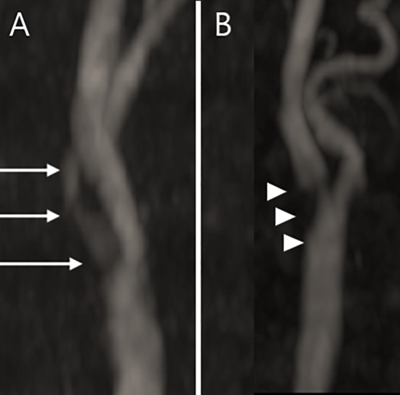
We retrospectively identified consecutive patients with acute symptomatic carotid disease who were admitted within 12 h after stroke onset. Fifty-nine patients underwent TOF MRA at admission and were categorized according to the presence or absence of intraplaque high signal intensity (HSI). The severity of carotid stenosis and diffusion-weighted magnetic resonance imaging lesion patterns were evaluated.
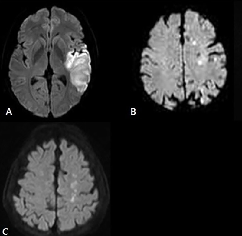
Intraplaque HSI was detected in 28.8% of the enrolled patients (17/59). Mild-to-moderate symptomatic carotid stenosis was more frequent in the intraplaque HSI-positive group (70.6%) than in the intraplaque HSI-negative group (42.8%) (p = 0.015). The patients with intraplaque HSI more frequently exhibited a disseminated small infarction pattern (76.5% in the intraplaque HSI-positive group, 47.6% in the -negative group), and did not exhibit a border-zone infarction pattern (0% in the positive group, 16.7% in the negative group).
TOF MRA may be a useful noninvasive and rapid tool to detect IPH in patients with acute symptomatic carotid disease. IPH was common in those with a lower degree of carotid stenosis and manifested as a disseminated small infarction pattern. Intraplaque HSI on TOF MRA in acute symptomatic carotid disease may help to determine the mechanism of stroke and establish early treatment plans.
颈动脉斑块内出血(IPH)是血栓栓塞的已知危险因素,但在急性症状性颈动脉疾病中快速检测 IPH 并不容易。本研究旨在评估时间飞跃(TOF)磁共振血管造影(MRA)在检测 IPH 中的效用,并评估急性症状性颈动脉疾病患者的狭窄程度和卒中模式。
我们回顾性地确定了在卒中发病后 12 小时内入院的连续急性症状性颈动脉疾病患者。59 例患者在入院时接受了 TOF MRA,并根据斑块内高信号强度(HSI)的存在与否进行分类。评估颈动脉狭窄的严重程度和弥散加权磁共振成像病变模式。
在纳入的患者中,有 28.8%(17/59)检测到斑块内 HSI。斑块内 HSI 阳性组(70.6%)中轻度至中度症状性颈动脉狭窄更为常见,而斑块内 HSI 阴性组(42.8%)则较少见(p=0.015)。斑块内 HSI 阳性组更频繁地表现出弥散性小梗死模式(76.5%,而斑块内 HSI 阴性组为 47.6%),而不表现出交界区梗死模式(阳性组为 0%,阴性组为 16.7%)。
TOF MRA 可能是一种有用的非侵入性快速工具,可用于检测急性症状性颈动脉疾病患者的 IPH。IPH 在颈动脉狭窄程度较低的患者中较为常见,表现为弥散性小梗死模式。急性症状性颈动脉疾病中 TOF MRA 的斑块内 HSI 可能有助于确定卒中的机制,并制定早期治疗计划。