• 文献检索
  • 文档翻译
  • 深度研究
  • 学术资讯
  • Suppr Zotero 插件Zotero 插件
  • 邀请有礼
  • 套餐&价格
  • 历史记录
应用&插件
Suppr Zotero 插件Zotero 插件浏览器插件Mac 客户端Windows 客户端微信小程序
定价
高级版会员购买积分包购买API积分包
服务
文献检索文档翻译深度研究API 文档MCP 服务
关于我们
关于 Suppr公司介绍联系我们用户协议隐私条款
关注我们

Suppr 超能文献

核心技术专利:CN118964589B侵权必究
粤ICP备2023148730 号-1Suppr @ 2026

文献检索

告别复杂PubMed语法,用中文像聊天一样搜索,搜遍4000万医学文献。AI智能推荐,让科研检索更轻松。

立即免费搜索

文件翻译

保留排版,准确专业,支持PDF/Word/PPT等文件格式,支持 12+语言互译。

免费翻译文档

深度研究

AI帮你快速写综述,25分钟生成高质量综述,智能提取关键信息,辅助科研写作。

立即免费体验

使用机器学习和定量相位成像技术对癌细胞的上皮和间充质特性进行定量评分。

Quantitative scoring of epithelial and mesenchymal qualities of cancer cells using machine learning and quantitative phase imaging.

机构信息

The Catholic Univ. of America, United States.

出版信息

J Biomed Opt. 2020 Feb;25(2):1-17. doi: 10.1117/1.JBO.25.2.026002.

DOI:10.1117/1.JBO.25.2.026002
PMID:32072775
原文链接:https://pmc.ncbi.nlm.nih.gov/articles/PMC7026523/
Abstract

SIGNIFICANCE

We introduce an application of machine learning trained on optical phase features of epithelial and mesenchymal cells to grade cancer cells' morphologies, relevant to evaluation of cancer phenotype in screening assays and clinical biopsies.

AIM

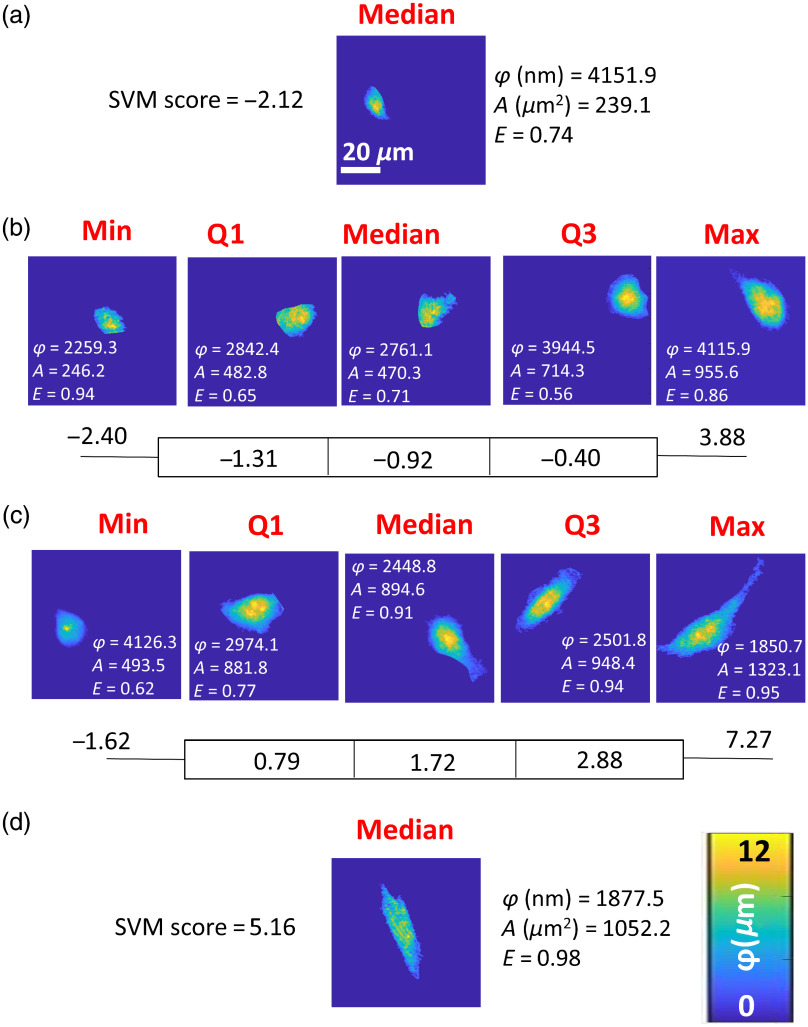
Our objective was to determine quantitative epithelial and mesenchymal qualities of breast cancer cells through an unbiased, generalizable, and linear score covering the range of observed morphologies.

APPROACH

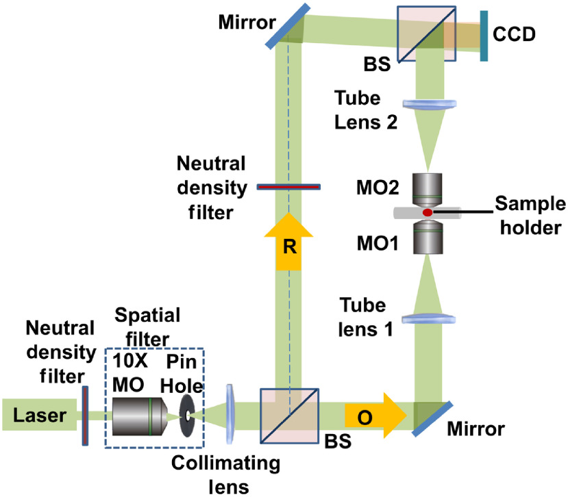
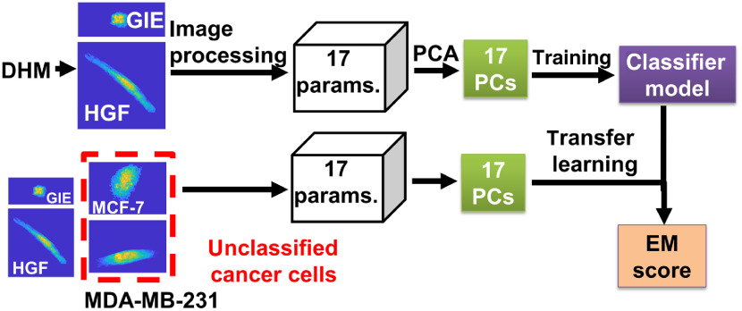
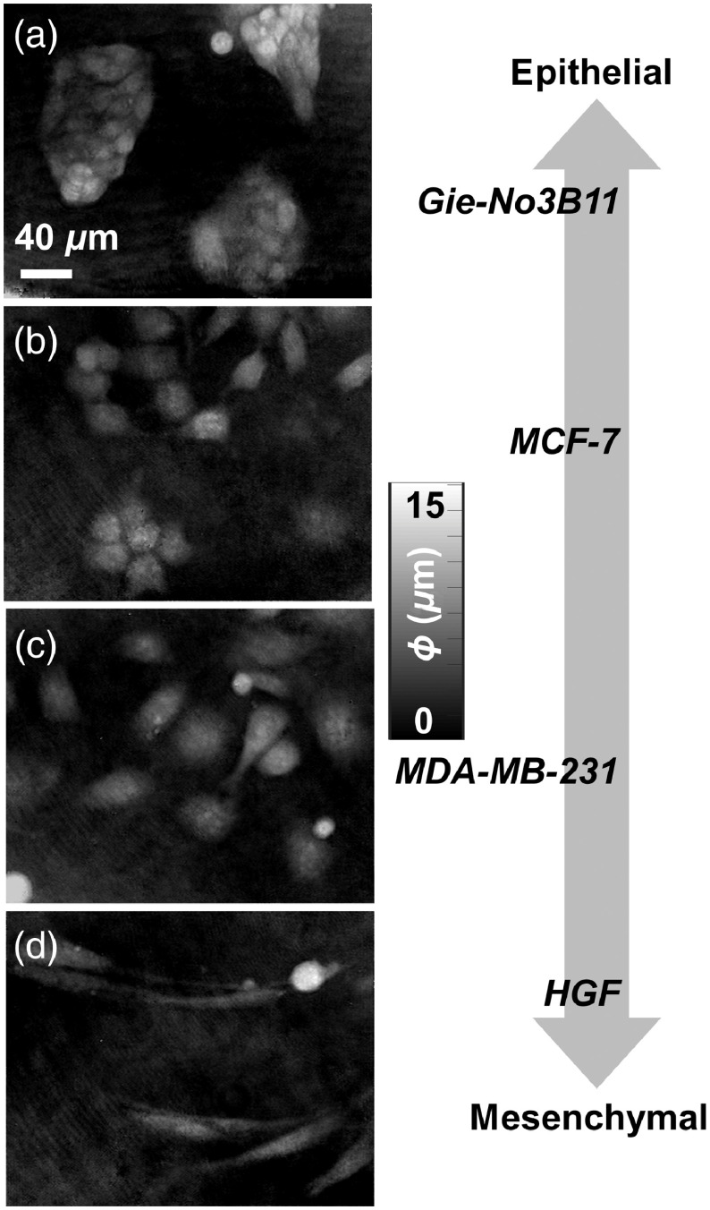
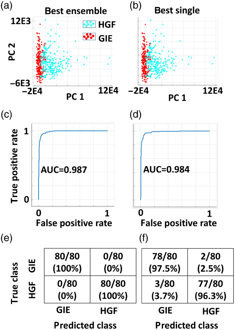
Digital holographic microscopy was used to generate phase height maps of noncancerous epithelial (Gie-No3B11) and fibroblast (human gingival) cell lines, as well as MDA-MB-231 and MCF-7 breast cancer cell lines. Several machine learning algorithms were evaluated as binary classifiers of the noncancerous cells that graded the cancer cells by transfer learning.

RESULTS

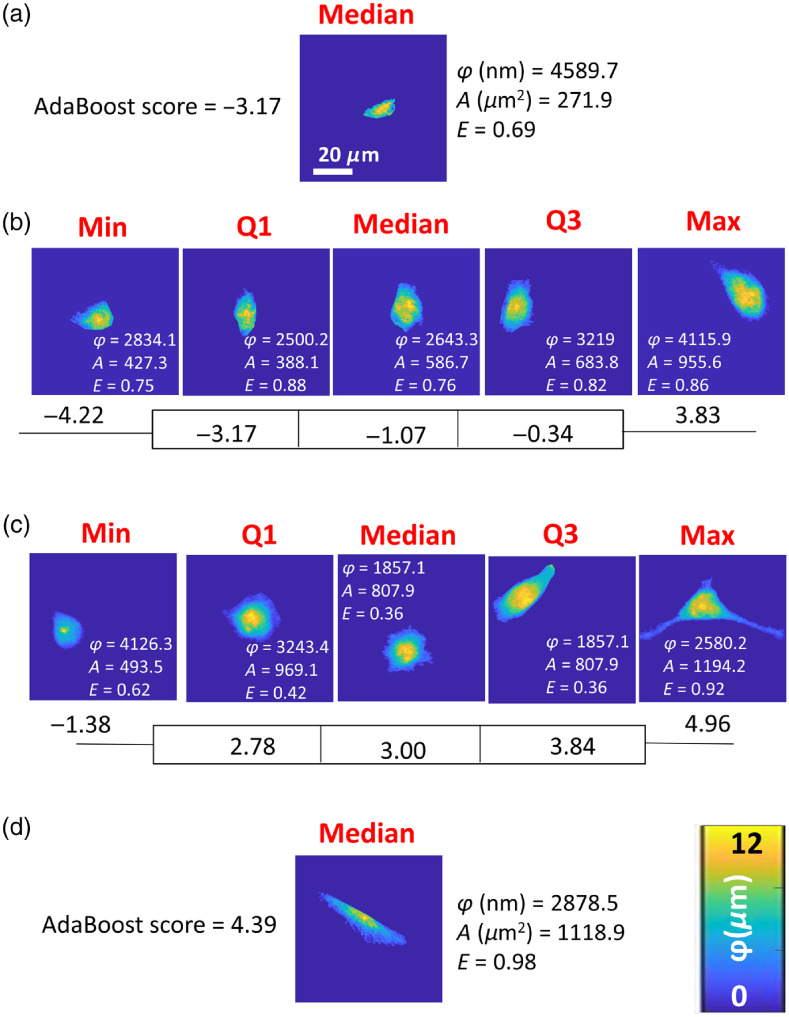
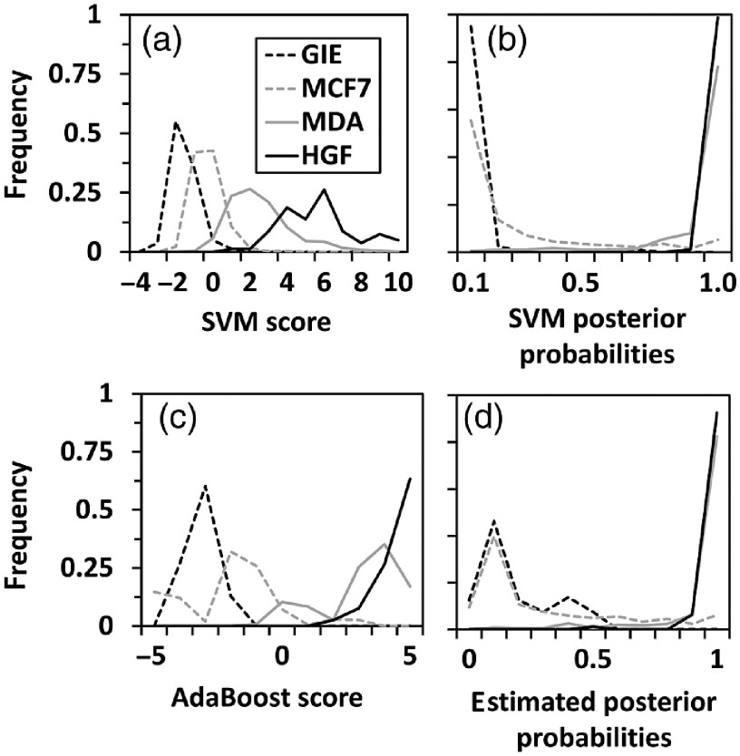
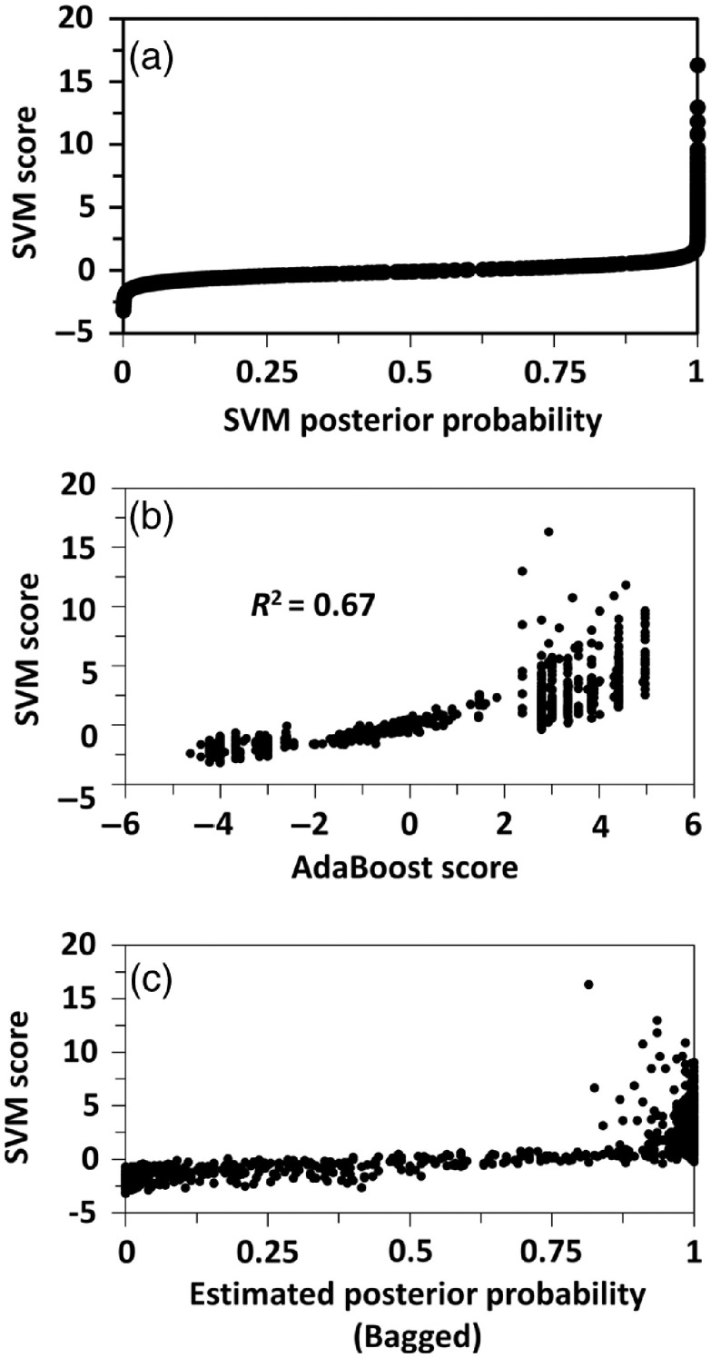
Epithelial and mesenchymal cells were classified with 96% to 100% accuracy. Breast cancer cells had scores in between the noncancer scores, indicating both epithelial and mesenchymal morphological qualities. The MCF-7 cells skewed toward epithelial scores, while MDA-MB-231 cells skewed toward mesenchymal scores. Linear support vector machines (SVMs) produced the most distinct score distributions for each cell line.

CONCLUSIONS

The proposed epithelial-mesenchymal score, derived from linear SVM learning, is a sensitive and quantitative approach for detecting epithelial and mesenchymal characteristics of unknown cells based on well-characterized cell lines. We establish a framework for rapid and accurate morphological evaluation of single cells and subtle phenotypic shifts in imaged cell populations.

摘要

意义

我们介绍了一种应用机器学习对上皮细胞和间充质细胞的光学相位特征进行训练,以对癌细胞的形态进行分级,这与筛选试验和临床活检中评估癌症表型相关。

目的

我们的目的是通过一个涵盖观察到的形态范围的无偏、可推广和线性评分,确定乳腺癌细胞的定量上皮和间充质特性。

方法

数字全息显微镜用于生成非癌上皮(Gie-No3B11)和成纤维细胞(人牙龈)细胞系以及 MDA-MB-231 和 MCF-7 乳腺癌细胞系的相位高度图。评估了几种机器学习算法作为非癌细胞的二进制分类器,通过迁移学习对癌细胞进行分级。

结果

上皮细胞和间充质细胞的分类准确率达到 96%至 100%。乳腺癌细胞的评分介于非癌评分之间,表明具有上皮和间充质形态特征。MCF-7 细胞偏向于上皮评分,而 MDA-MB-231 细胞偏向于间充质评分。线性支持向量机(SVM)产生了每个细胞系最独特的评分分布。

结论

基于线性 SVM 学习得出的拟议上皮-间充质评分是一种敏感和定量的方法,可根据特征明确的细胞系检测未知细胞的上皮和间充质特征。我们建立了一个快速准确评估单细胞形态和成像细胞群体中细微表型变化的框架。

https://cdn.ncbi.nlm.nih.gov/pmc/blobs/d1f6/7026523/c089761c5a62/JBO-025-026002-g008.jpg
https://cdn.ncbi.nlm.nih.gov/pmc/blobs/d1f6/7026523/a20407a1388a/JBO-025-026002-g001.jpg
https://cdn.ncbi.nlm.nih.gov/pmc/blobs/d1f6/7026523/fd6a0df146ba/JBO-025-026002-g002.jpg
https://cdn.ncbi.nlm.nih.gov/pmc/blobs/d1f6/7026523/557641c77453/JBO-025-026002-g003.jpg
https://cdn.ncbi.nlm.nih.gov/pmc/blobs/d1f6/7026523/c4589820db8e/JBO-025-026002-g004.jpg
https://cdn.ncbi.nlm.nih.gov/pmc/blobs/d1f6/7026523/b98a942eba2e/JBO-025-026002-g005.jpg
https://cdn.ncbi.nlm.nih.gov/pmc/blobs/d1f6/7026523/0a912035a043/JBO-025-026002-g006.jpg
https://cdn.ncbi.nlm.nih.gov/pmc/blobs/d1f6/7026523/0a7b71bdfa9c/JBO-025-026002-g007.jpg
https://cdn.ncbi.nlm.nih.gov/pmc/blobs/d1f6/7026523/c089761c5a62/JBO-025-026002-g008.jpg

相似文献

1
Quantitative scoring of epithelial and mesenchymal qualities of cancer cells using machine learning and quantitative phase imaging.使用机器学习和定量相位成像技术对癌细胞的上皮和间充质特性进行定量评分。
J Biomed Opt. 2020 Feb;25(2):1-17. doi: 10.1117/1.JBO.25.2.026002.
2
Quantitative assessment of cancer cell morphology and motility using telecentric digital holographic microscopy and machine learning.利用远心数字全息显微镜和机器学习对癌细胞形态和运动进行定量评估。
Cytometry A. 2018 Mar;93(3):334-345. doi: 10.1002/cyto.a.23316. Epub 2017 Dec 28.
3
Machine Learning with Optical Phase Signatures for Phenotypic Profiling of Cell Lines.基于光学相位特征的机器学习在细胞系表型分析中的应用。
Cytometry A. 2019 Jul;95(7):757-768. doi: 10.1002/cyto.a.23774. Epub 2019 Apr 22.
4
Automated interpretation of time-lapse quantitative phase image by machine learning to study cellular dynamics during epithelial-mesenchymal transition.基于机器学习的时间 lapse 定量相位图像自动解释,用于研究上皮间质转化过程中的细胞动力学。
J Biomed Opt. 2020 Aug;25(8). doi: 10.1117/1.JBO.25.8.086502.
5
TOP-GAN: Stain-free cancer cell classification using deep learning with a small training set.TOP-GAN:使用深度学习和小训练集进行无染色癌细胞分类
Med Image Anal. 2019 Oct;57:176-185. doi: 10.1016/j.media.2019.06.014. Epub 2019 Jun 26.
6
Automated classification of cell morphology by coherence-controlled holographic microscopy.通过相干控制全息显微镜实现细胞形态的自动分类
J Biomed Opt. 2017 Aug;22(8):1-9. doi: 10.1117/1.JBO.22.8.086008.
7
Quantitative phase microscopy spatial signatures of cancer cells.癌细胞的定量相显微镜空间特征。
Cytometry A. 2017 May;91(5):482-493. doi: 10.1002/cyto.a.23100. Epub 2017 Apr 20.
8
Digital holographic technique based breast cancer detection using transfer learning method.基于迁移学习方法的数字全息乳腺癌检测技术。
J Biophotonics. 2023 Aug;16(8):e202200359. doi: 10.1002/jbio.202200359. Epub 2023 May 2.
9
Macrophage phenotypic subtypes diametrically regulate epithelial-mesenchymal plasticity in breast cancer cells.巨噬细胞表型亚型对乳腺癌细胞的上皮-间质可塑性起着截然相反的调节作用。
BMC Cancer. 2016 Jul 7;16:419. doi: 10.1186/s12885-016-2411-1.
10
Lung epithelial cells induce both phenotype alteration and senescence in breast cancer cells.肺上皮细胞可诱导乳腺癌细胞表型改变和衰老。
PLoS One. 2015 Jan 30;10(1):e0118060. doi: 10.1371/journal.pone.0118060. eCollection 2015.

引用本文的文献

1
Morphological and Optical Profiling of Melanocytes and SK-MEL-28 Melanoma Cells Via Digital Holographic Microscopy and Quantitative Phase Imaging.通过数字全息显微镜和定量相成像对黑素细胞和SK-MEL-28黑色素瘤细胞进行形态学和光学分析。
Adv Biol (Weinh). 2025 Feb;9(2):e2400346. doi: 10.1002/adbi.202400346. Epub 2024 Nov 11.
2
Rapid mechanical phenotyping of breast cancer cells based on stochastic intracellular fluctuations.基于随机细胞内波动的乳腺癌细胞快速机械表型分析
iScience. 2024 Oct 4;27(11):110960. doi: 10.1016/j.isci.2024.110960. eCollection 2024 Nov 15.
3
Grading of glioma tumors using digital holographic microscopy.

本文引用的文献

1
Quantitative assessment of changes in cellular morphology at photodynamic treatment by means of digital holographic microscopy.通过数字全息显微镜对光动力治疗时细胞形态变化进行定量评估。
Biomed Opt Express. 2019 Sep 6;10(10):4975-4986. doi: 10.1364/BOE.10.004975. eCollection 2019 Oct 1.
2
TOP-GAN: Stain-free cancer cell classification using deep learning with a small training set.TOP-GAN:使用深度学习和小训练集进行无染色癌细胞分类
Med Image Anal. 2019 Oct;57:176-185. doi: 10.1016/j.media.2019.06.014. Epub 2019 Jun 26.
3
Quantitative phase imaging of cells in a flow cytometry arrangement utilizing Michelson interferometer-based off-axis digital holographic microscopy.
使用数字全息显微镜对胶质瘤肿瘤进行分级。
Heliyon. 2024 Apr 23;10(9):e29897. doi: 10.1016/j.heliyon.2024.e29897. eCollection 2024 May 15.
4
On the use of deep learning for phase recovery.关于深度学习在相位恢复中的应用。
Light Sci Appl. 2024 Jan 1;13(1):4. doi: 10.1038/s41377-023-01340-x.
5
Artificial intelligence-enabled quantitative phase imaging methods for life sciences.人工智能赋能的生命科学定量相位成像方法。
Nat Methods. 2023 Nov;20(11):1645-1660. doi: 10.1038/s41592-023-02041-4. Epub 2023 Oct 23.
6
Digital Holographic Microscopy to Assess Cell Behavior.数字全息显微镜评估细胞行为。
Methods Mol Biol. 2023;2644:247-266. doi: 10.1007/978-1-0716-3052-5_16.
7
Long-term label-free assessments of individual bacteria using three-dimensional quantitative phase imaging and hydrogel-based immobilization.使用三维定量相衬成像和水凝胶固定化技术对单个细菌进行长期无标记评估。
Sci Rep. 2023 Jan 2;13(1):46. doi: 10.1038/s41598-022-27158-y.
8
Quantitative Phase Imaging: Recent Advances and Expanding Potential in Biomedicine.定量相位成像:生物医学中的最新进展和扩展潜力。
ACS Nano. 2022 Aug 23;16(8):11516-11544. doi: 10.1021/acsnano.1c11507. Epub 2022 Aug 2.
9
Cancer-Cell Deep-Learning Classification by Integrating Quantitative-Phase Spatial and Temporal Fluctuations.基于定量相位空间和时间波动的癌细胞深度学习分类。
Cells. 2021 Nov 29;10(12):3353. doi: 10.3390/cells10123353.
10
Label-Free and Quantitative Dry Mass Monitoring for Single Cells during In Situ Culture.无标记和定量干物质监测单细胞在原位培养过程中。
Cells. 2021 Jun 29;10(7):1635. doi: 10.3390/cells10071635.
利用基于迈克尔逊干涉仪的离轴数字全息显微镜在流式细胞仪配置中对细胞进行定量相成像。
J Biophotonics. 2019 Sep;12(9):e201900085. doi: 10.1002/jbio.201900085. Epub 2019 Jun 20.
4
Quantitative Phase Imaging Flow Cytometry for Ultra-Large-Scale Single-Cell Biophysical Phenotyping.用于超大规模单细胞生物物理表型分析的定量相位成像流式细胞术。
Cytometry A. 2019 May;95(5):510-520. doi: 10.1002/cyto.a.23765. Epub 2019 Apr 22.
5
Machine Learning with Optical Phase Signatures for Phenotypic Profiling of Cell Lines.基于光学相位特征的机器学习在细胞系表型分析中的应用。
Cytometry A. 2019 Jul;95(7):757-768. doi: 10.1002/cyto.a.23774. Epub 2019 Apr 22.
6
Unsupervised organization of cervical cells using bright-field and single-shot digital holographic microscopy.使用明场和单次数字全息显微镜对宫颈细胞进行无监督组织分析。
J Biophotonics. 2019 Aug;12(8):e201800409. doi: 10.1002/jbio.201800409. Epub 2019 May 2.
7
An optical study of drug resistance detection in endometrial cancer cells by dynamic and quantitative phase imaging.动态和定量相位成像在子宫内膜癌细胞耐药性检测中的光学研究。
J Biophotonics. 2019 Jul;12(7):e201800443. doi: 10.1002/jbio.201800443. Epub 2019 Apr 2.
8
PhaseStain: the digital staining of label-free quantitative phase microscopy images using deep learning.相位染色:使用深度学习对无标记定量相显微镜图像进行数字染色。
Light Sci Appl. 2019 Feb 6;8:23. doi: 10.1038/s41377-019-0129-y. eCollection 2019.
9
Multi-ATOM: Ultrahigh-throughput single-cell quantitative phase imaging with subcellular resolution.多原子:具有亚细胞分辨率的超高通量单细胞定量相位成像。
J Biophotonics. 2019 Jul;12(7):e201800479. doi: 10.1002/jbio.201800479. Epub 2019 Apr 1.
10
Label-free classification of cells based on supervised machine learning of subcellular structures.基于亚细胞结构的有监督机器学习对细胞进行无标记分类。
PLoS One. 2019 Jan 29;14(1):e0211347. doi: 10.1371/journal.pone.0211347. eCollection 2019.