Research Imaging Institute, University of Texas Health Science Center at San Antonio (Gray, Fox); Institute of Neuroscience and Medicine, Brain and Behavior (INM-7), Research Center Jüelich, Germany (Müller, Eickhoff); Institute of Systems Neuroscience, Medical Faculty, Heinrich Heine University Düsseldorf, Germany (Müller, Eickhoff); and South Texas Veterans Health Care System, San Antonio (Fox).
Am J Psychiatry. 2020 May 1;177(5):422-434. doi: 10.1176/appi.ajp.2019.19050560. Epub 2020 Feb 26.
Imaging studies of major depressive disorder have reported structural and functional abnormalities in a variety of spatially diverse brain regions. Quantitative meta-analyses of this literature, however, have failed to find statistically significant between-study spatial convergence, other than transdiagnostic-only effects. In the present study, the authors applied a novel multimodal meta-analytic approach to test the hypothesis that major depression exhibits spatially convergent structural and functional brain abnormalities.
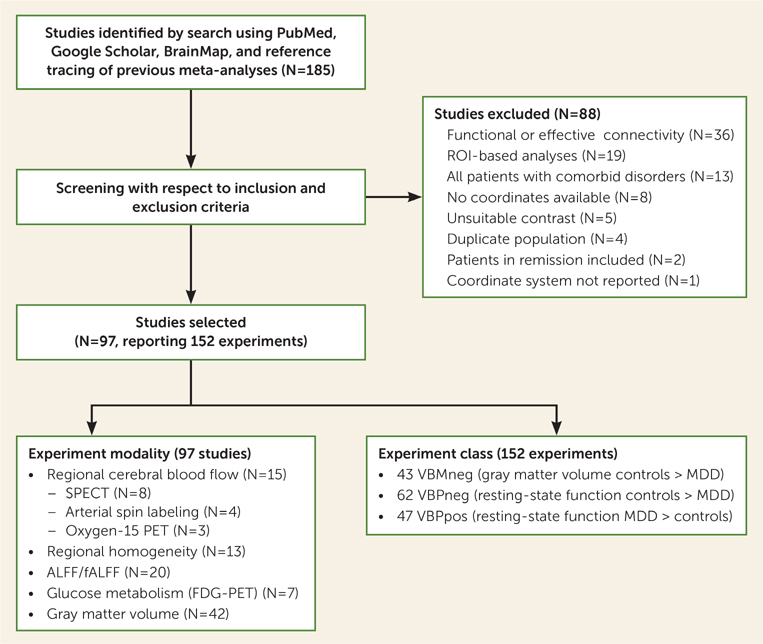
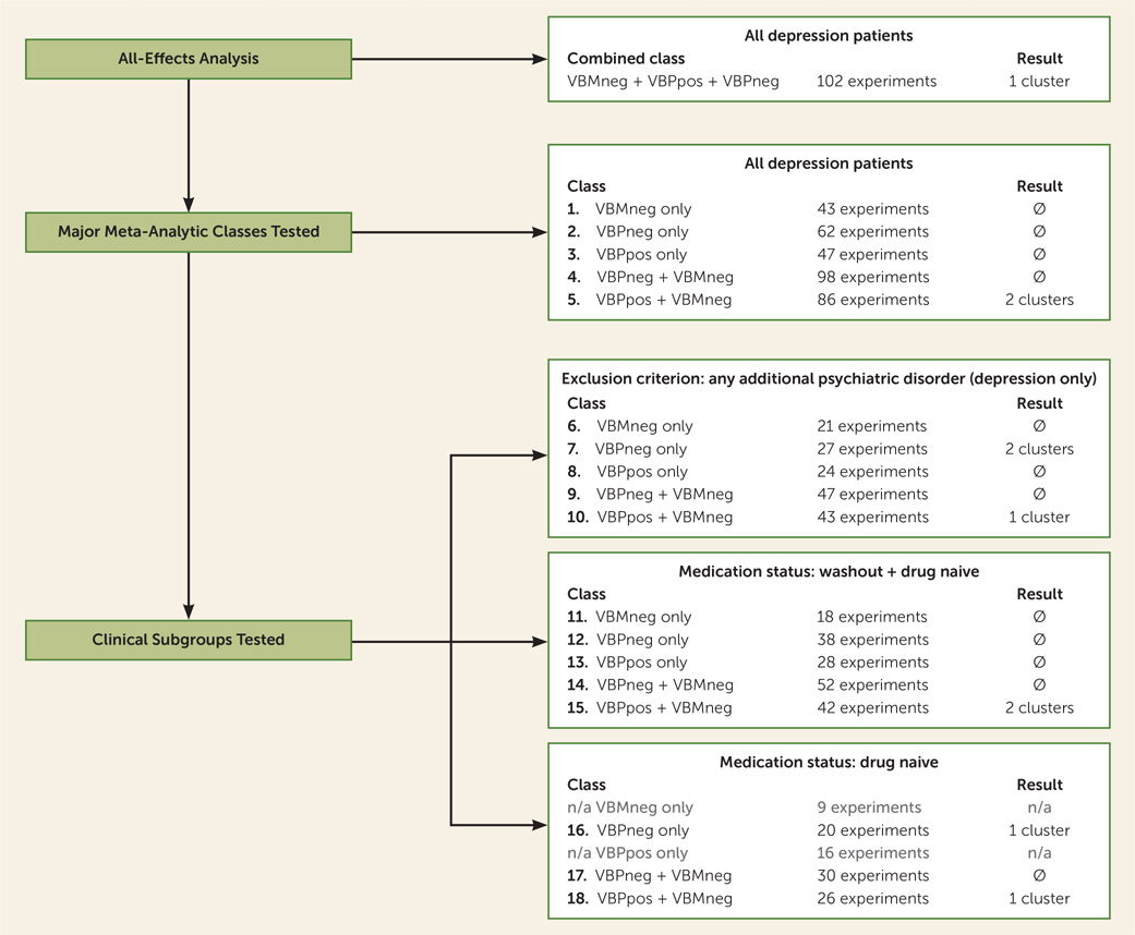
This coordinate-based meta-analysis included voxel-based morphometry (VBM) studies and resting-state voxel-based pathophysiology (VBP) studies of blood flow, glucose metabolism, regional homogeneity, and amplitude of low-frequency fluctuations (ALFF) and fractional ALFF (fALFF). Input data were grouped into three primary meta-analytic classes: gray matter atrophy, increased function, and decreased function in patients with major depression relative to healthy control subjects. In secondary meta-analyses, the data were grouped across primary categories, and in tertiary analyses, by medication status and absence of psychiatric comorbidity. Activation likelihood estimation was used for all analyses.
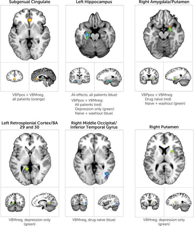
A total of 92 publications reporting 152 experiments were identified, collectively representing 2,928 patients with major depressive disorder. The primary analyses detected no convergence across studies. The secondary analyses identified portions of the subgenual cingulate cortex, hippocampus, amygdala, and putamen as demonstrating convergent abnormalities. The tertiary analyses (clinical subtypes) showed improved convergence relative to the secondary analyses.
Coordinate-based meta-analysis identified spatially convergent structural (VBM) and functional (VBP) abnormalities in major depression. The findings suggest replicable neuroimaging features associated with major depression, beyond the transdiagnostic effects reported in previous meta-analyses, and support a continued research focus on the subgenual cingulate and other selected regions' role in depression.
重度抑郁症的影像学研究报告称,各种空间上不同的大脑区域存在结构和功能异常。然而,对该文献的定量荟萃分析并未发现除跨诊断效应外的统计学上显著的研究间空间收敛性。在本研究中,作者应用了一种新的多模态荟萃分析方法来检验这样一个假设,即重度抑郁症表现出空间上一致的结构和功能脑异常。
该基于坐标的荟萃分析包括基于体素的形态测量学(VBM)研究和静息状态基于体素的病理生理学(VBP)研究,用于血流、葡萄糖代谢、局部一致性以及低频波动的幅度(ALFF)和分数 ALFF(fALFF)。输入数据分为三个主要的荟萃分析类别:与健康对照组相比,重度抑郁症患者的灰质萎缩、功能增强和功能减退。在二级荟萃分析中,数据按主要类别分组,在三级分析中,按药物状态和无精神共病分组。所有分析均使用激活似然估计法。
共确定了 92 篇报告 152 项实验的出版物,共代表了 2928 例重度抑郁症患者。主要分析未发现研究间的收敛性。二级分析确定了扣带回皮质下、海马体、杏仁核和壳核的某些部分存在一致的异常。三级分析(临床亚型)显示与二级分析相比,收敛性得到改善。
基于坐标的荟萃分析确定了重度抑郁症存在空间一致的结构(VBM)和功能(VBP)异常。这些发现表明存在与以前荟萃分析中报告的跨诊断效应不同的、与重度抑郁症相关的可复制神经影像学特征,并支持继续关注扣带回皮质下和其他选定区域在抑郁症中的作用。