Tharwat Mohamed
Department of Veterinary Medicine, College of Agriculture and Veterinary Medicine, Qassim University, P.O. Box 6622, Buraidah, 51452, Saudi Arabia.
Department of Animal Medicine, Faculty of Veterinary Medicine, Zagazig University, 44519, Zagazig, Egypt.
J Vet Med Sci. 2020 Apr 9;82(4):399-407. doi: 10.1292/jvms.19-0690. Epub 2020 Feb 26.
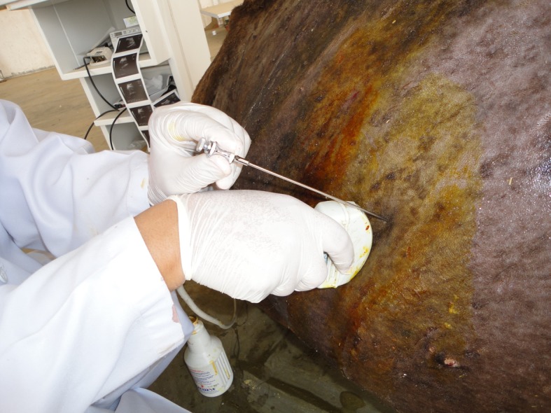
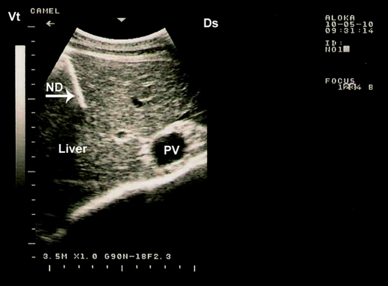
In camels, hepatic diseases are relatively common and most of them are misdiagnosed as a cause of illness because signs may be subtle. In addition, diagnostic laboratory methods are insufficient as hepatic enzymes can also be elevated in camels with cardiac or skeletal muscle damage. Examples of liver diseases in camels are hepatic lipidosis, hepatitis, cirrhosis, hepatic necrosis, choleostasis, hyperplasia of biliary epithelium, hydatid cysts, glycogen deposition, cholangitis, cholangiohepatitis, calcified hydatid cyst and hepatic abscesses. When the liver is examined by ultrasonography, the clinician gets sufficient information about the size, position, echopatterns of the hepatic parenchyma, bile ducts and outlines of the hepatic blood vessels. Ultrasonography has been used previously in camels only for reproductive purposes. However, during the past decade, it has been used for scanning of the healthy organs as well as evaluation and determining the diagnosis and prognosis of non-reproductive disorders. Examples of diseases evaluated by ultrasonography in camels are paratuberculosis, trypanosomiasis, abdominal and urinary disorders, thoracic diseases, renal tumors, pyelonephritis, renal abscessation, gastrointestinal tumors, chronic peritonitis and splenic abscessation. Ultrasound-guidance in biopsy of hepatic lesions and in portocentesis has also been reported in camels. This mini review article is written to shed light on ultrasonography of the liver and its blood vessels in healthy camels as well as finding in camels with hepatic disorders such as fatty infiltration of the liver, hepatic abscesses and calcification of the bile ducts.
在骆驼中,肝脏疾病相对常见,而且由于症状可能不明显,大多数此类疾病会被误诊为致病原因。此外,诊断性实验室方法并不充分,因为患有心脏或骨骼肌损伤的骆驼体内肝酶也可能升高。骆驼肝脏疾病的例子包括肝脂肪变性、肝炎、肝硬化、肝坏死、胆汁淤积、胆管上皮增生、包虫囊肿、糖原沉积、胆管炎、胆管肝炎、钙化包虫囊肿和肝脓肿。当通过超声检查肝脏时,临床医生可以获得有关肝脏实质、胆管和肝血管轮廓的大小、位置、回声模式的充分信息。超声检查以前在骆驼中仅用于生殖目的。然而,在过去十年中,它也被用于扫描健康器官以及评估和确定非生殖系统疾病的诊断和预后。通过超声检查评估骆驼疾病的例子包括副结核病、锥虫病、腹部和泌尿系统疾病、胸部疾病、肾肿瘤、肾盂肾炎、肾脓肿、胃肠道肿瘤、慢性腹膜炎和脾脓肿。在骆驼中也有关于肝脏病变活检和门静脉穿刺超声引导的报道。这篇小型综述文章旨在阐明健康骆驼肝脏及其血管的超声检查情况,以及患有肝脏疾病(如肝脏脂肪浸润、肝脓肿和胆管钙化)骆驼的检查结果。