Institute of Medical Engineering, Department of Biophysics, School of Basic Medical Science, Health Science Center, Xi'an Jiaotong University, No.76 Yanta West Road, Xi'an, Shaanxi 710061, PR China.; Department of Radiology, Weill Cornell Medicine, New York, NY 10065, USA.
Department of Radiology, Weill Cornell Medicine, New York, NY 10065, USA.; Department of Gastrointestinal Surgery, The Second Clinical Medicine College (Shenzhen People's Hospital) of Jinan University, Shenzhen, Guangdong 518020, China.
Int J Biol Macromol. 2020 Jun 15;153:100-106. doi: 10.1016/j.ijbiomac.2020.02.253. Epub 2020 Feb 24.
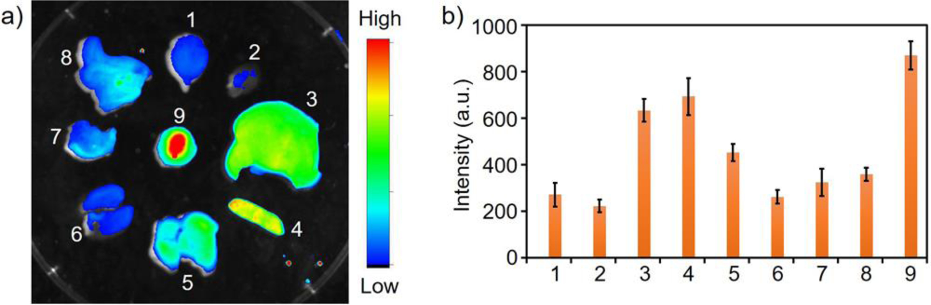
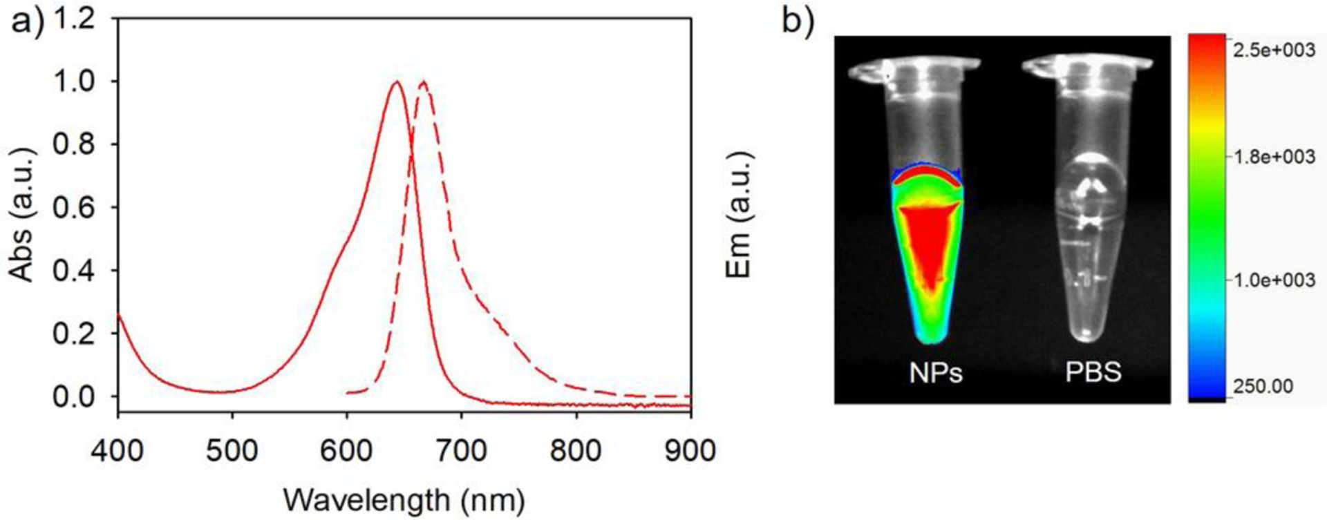
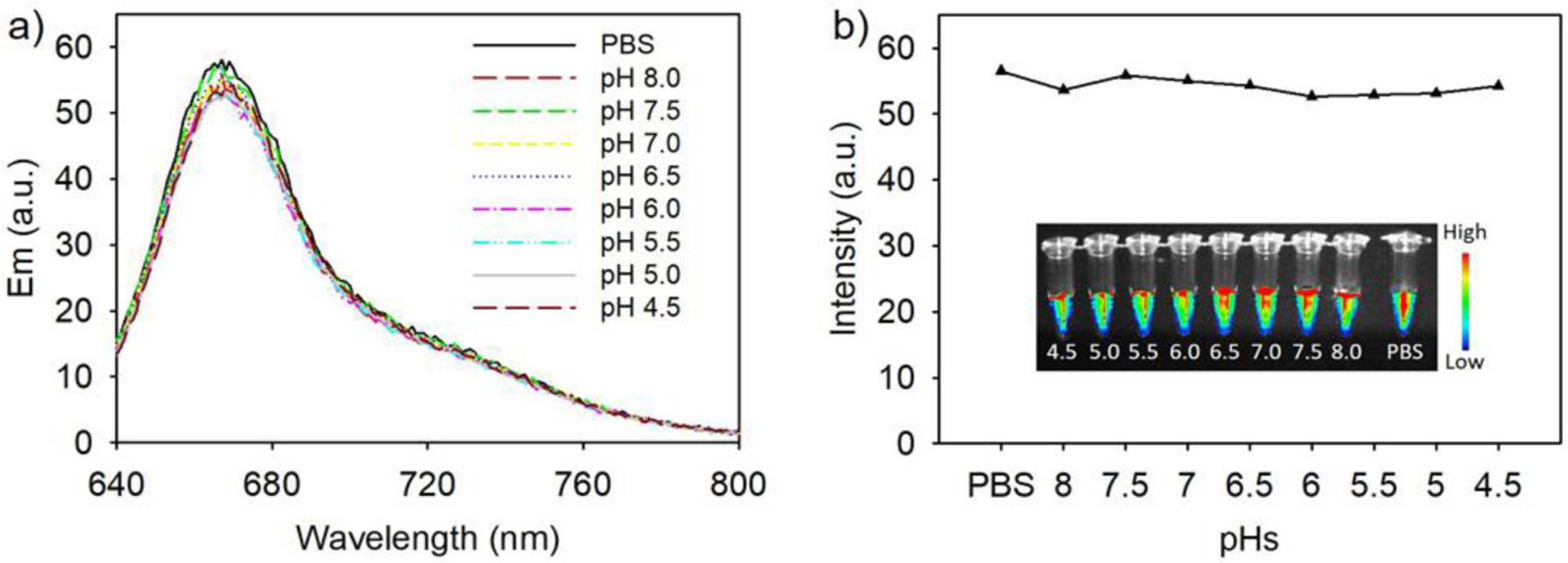
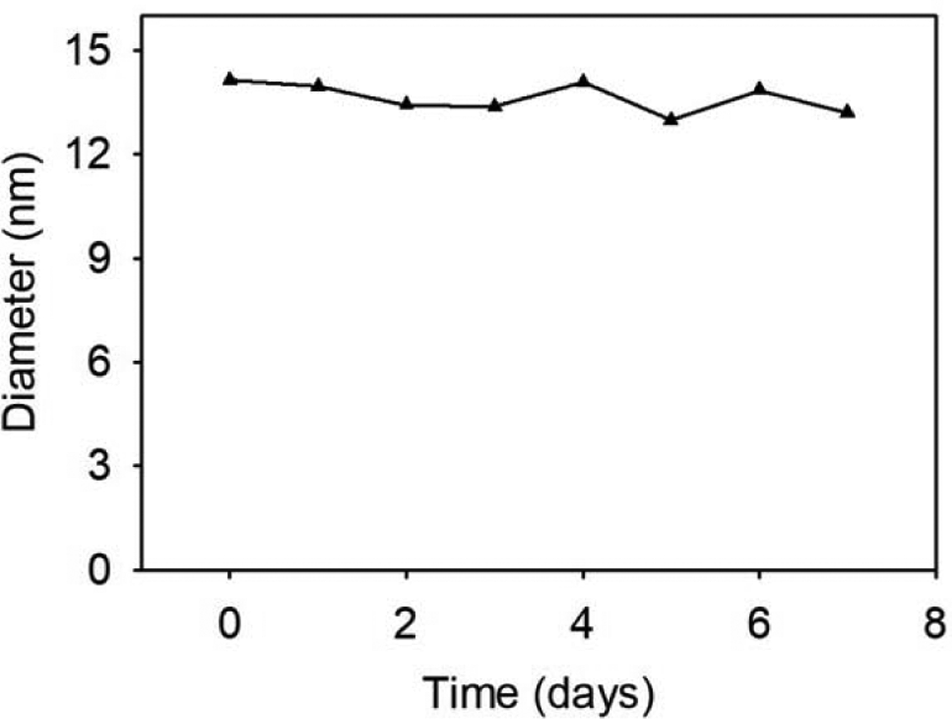
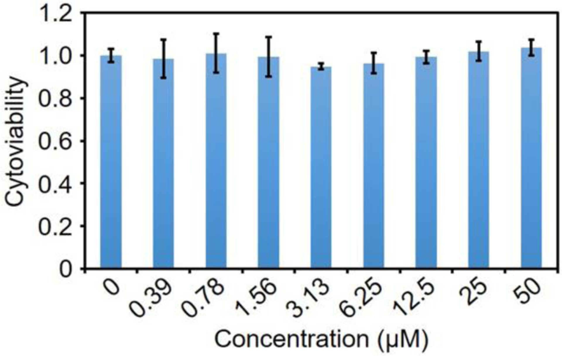
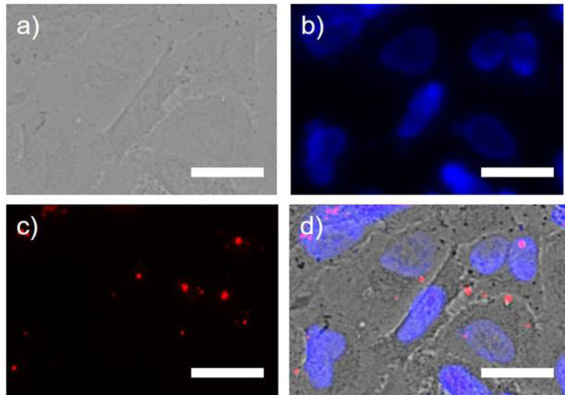
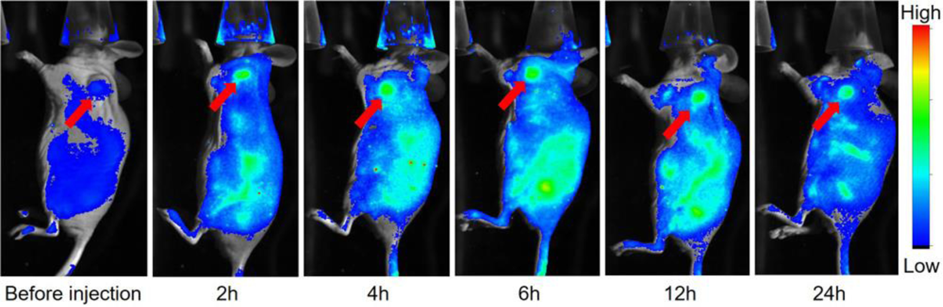
Nanoparticles are excellent imaging agents for cancer, but variability in chemical structure, racemic mixtures, and addition of heavy metals hinders FDA approval in the United States. We developed a small ultra-red fluorescent protein, named smURFP, to have optical properties similar to the small-molecule Cy5, a heptamethine subclass of cyanine dyes (Ex/Em = 642/670 nm). smURFP has a fluorescence quantum yield of 18% and expresses so well in E. coli, that gram quantities of fluorescent protein are purified from cultures in the laboratory. In this research, the fluorescent protein smURFP was combined with bovine serum albumin into fluorescent protein nanoparticles. These nanoparticles are fluorescent with a quantum yield of 17% and 12-14 nm in diameter. The far-red fluorescent protein nanoparticles noninvasively image tumors in living mice via the enhanced permeation and retention (EPR) mechanism. This manuscript describes the use of a new fluorescent protein nanoparticle for in vivo fluorescent imaging. This protein nanoparticle core should prove useful as a biomacromolecular scaffold, which could bear extended chemical modifications for studies, such as the in vivo imaging of fluorescent protein nanoparticles targeted to primary and metastatic cancer, theranostic treatment, and/or dual-modality imaging with positron emission tomography for entire human imaging.
纳米颗粒是癌症的优秀成像剂,但化学结构的可变性、外消旋混合物和重金属的添加阻碍了它们在美国获得 FDA 的批准。我们开发了一种小型超红色荧光蛋白,命名为 smURFP,其光学性质与小分子 Cy5 相似,Cy5 是一种菁染料的七甲川类(Ex/Em = 642/670nm)。smURFP 的荧光量子产率为 18%,在大肠杆菌中表达良好,以至于可以从实验室培养物中纯化出克级别的荧光蛋白。在这项研究中,将荧光蛋白 smURFP 与牛血清白蛋白结合成荧光蛋白纳米颗粒。这些纳米颗粒具有荧光特性,其荧光量子产率为 17%,直径为 12-14nm。远红荧光蛋白纳米颗粒通过增强渗透和保留(EPR)机制非侵入性地对活体小鼠中的肿瘤进行成像。本文描述了使用新型荧光蛋白纳米颗粒进行体内荧光成像。这种蛋白纳米颗粒核心有望成为一种生物大分子支架,它可以进行扩展的化学修饰,用于研究,例如针对原发性和转移性癌症的荧光蛋白纳米颗粒的体内成像、治疗和/或与正电子发射断层扫描的双模式成像整个人体成像。