• 文献检索
  • 文档翻译
  • 深度研究
  • 学术资讯
  • Suppr Zotero 插件Zotero 插件
  • 邀请有礼
  • 套餐&价格
  • 历史记录
应用&插件
Suppr Zotero 插件Zotero 插件浏览器插件Mac 客户端Windows 客户端微信小程序
定价
高级版会员购买积分包购买API积分包
服务
文献检索文档翻译深度研究API 文档MCP 服务
关于我们
关于 Suppr公司介绍联系我们用户协议隐私条款
关注我们

Suppr 超能文献

核心技术专利:CN118964589B侵权必究
粤ICP备2023148730 号-1Suppr @ 2026

文献检索

告别复杂PubMed语法,用中文像聊天一样搜索,搜遍4000万医学文献。AI智能推荐,让科研检索更轻松。

立即免费搜索

文件翻译

保留排版,准确专业,支持PDF/Word/PPT等文件格式,支持 12+语言互译。

免费翻译文档

深度研究

AI帮你快速写综述,25分钟生成高质量综述,智能提取关键信息,辅助科研写作。

立即免费体验

高级别胶质瘤瘤内异质性的影像学表现。

Imaging of intratumoral heterogeneity in high-grade glioma.

机构信息

Department of Radiology, Mayo Clinic Arizona, 5777 E Mayo Blvd, Phoenix, AZ, 85054, USA.

Mathematical NeuroOncology Lab, Precision Neurotherapeutics Innovation Program, Mayo Clinic, 5777 East Mayo Blvd, Support, Services Building Suite 2-700, Phoenix, AZ, 85054, USA.

出版信息

Cancer Lett. 2020 May 1;477:97-106. doi: 10.1016/j.canlet.2020.02.025. Epub 2020 Feb 27.

DOI:10.1016/j.canlet.2020.02.025
PMID:32112907
原文链接:https://pmc.ncbi.nlm.nih.gov/articles/PMC7108976/
Abstract

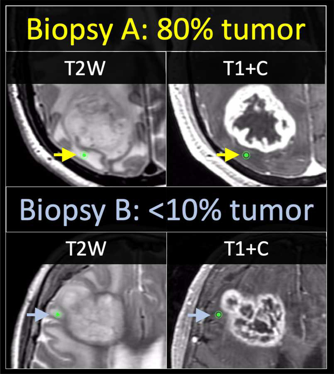
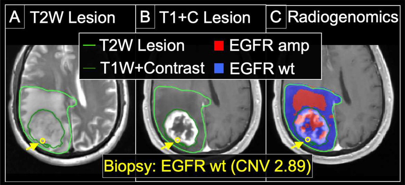
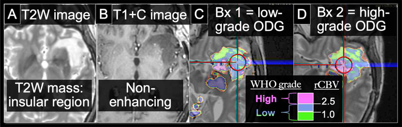
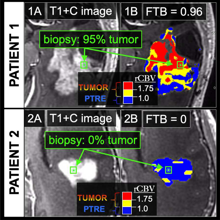
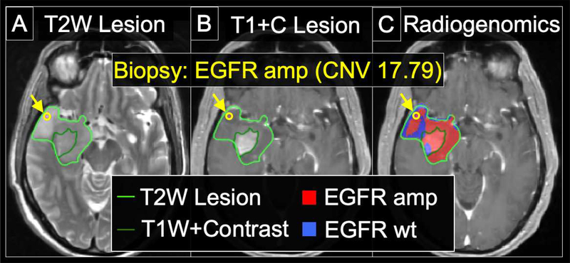
High-grade glioma (HGG), and particularly Glioblastoma (GBM), can exhibit pronounced intratumoral heterogeneity that confounds clinical diagnosis and management. While conventional contrast-enhanced MRI lacks the capability to resolve this heterogeneity, advanced MRI techniques and PET imaging offer a spectrum of physiologic and biophysical image features to improve the specificity of imaging diagnoses. Published studies have shown how integrating these advanced techniques can help better define histologically distinct targets for surgical and radiation treatment planning, and help evaluate the regional heterogeneity of tumor recurrence and response assessment following standard adjuvant therapy. Application of texture analysis and machine learning (ML) algorithms has also enabled the emerging field of radiogenomics, which can spatially resolve the regional and genetically distinct subpopulations that coexist within a single GBM tumor. This review focuses on the latest advances in neuro-oncologic imaging and their clinical applications for the assessment of intratumoral heterogeneity.

摘要

高级别胶质瘤(HGG),尤其是胶质母细胞瘤(GBM),可能表现出明显的肿瘤内异质性,这使得临床诊断和管理变得复杂。虽然常规的对比增强 MRI 缺乏解析这种异质性的能力,但先进的 MRI 技术和 PET 成像提供了一系列生理和生物物理的图像特征,以提高成像诊断的特异性。已发表的研究表明,如何整合这些先进技术可以帮助更好地定义手术和放射治疗计划中具有组织学差异的目标,并有助于评估标准辅助治疗后肿瘤复发和反应评估的区域异质性。纹理分析和机器学习(ML)算法的应用也使得放射基因组学这一新兴领域成为可能,该领域可以在单个 GBM 肿瘤内共存的具有空间分辨率的区域和遗传上不同的亚群。这篇综述重点介绍神经肿瘤学成像的最新进展及其在评估肿瘤内异质性方面的临床应用。

https://cdn.ncbi.nlm.nih.gov/pmc/blobs/c4f5/7108976/7531cc779e1e/nihms-1568647-f0005.jpg
https://cdn.ncbi.nlm.nih.gov/pmc/blobs/c4f5/7108976/f3f4225177a4/nihms-1568647-f0001.jpg
https://cdn.ncbi.nlm.nih.gov/pmc/blobs/c4f5/7108976/deac0ff75c42/nihms-1568647-f0002.jpg
https://cdn.ncbi.nlm.nih.gov/pmc/blobs/c4f5/7108976/57f4b86f7fb4/nihms-1568647-f0003.jpg
https://cdn.ncbi.nlm.nih.gov/pmc/blobs/c4f5/7108976/861f2a8985a1/nihms-1568647-f0004.jpg
https://cdn.ncbi.nlm.nih.gov/pmc/blobs/c4f5/7108976/7531cc779e1e/nihms-1568647-f0005.jpg

相似文献

1
Imaging of intratumoral heterogeneity in high-grade glioma.高级别胶质瘤瘤内异质性的影像学表现。
Cancer Lett. 2020 May 1;477:97-106. doi: 10.1016/j.canlet.2020.02.025. Epub 2020 Feb 27.
2
High-grade glioma radiation therapy target volumes and patterns of failure obtained from magnetic resonance imaging and 18F-FDOPA positron emission tomography delineations from multiple observers.从多位观察者的磁共振成像和 18F-FDOPA 正电子发射断层扫描勾画中获得高级别胶质瘤放射治疗靶区和失败模式。
Int J Radiat Oncol Biol Phys. 2013 Dec 1;87(5):1100-6. doi: 10.1016/j.ijrobp.2013.09.008. Epub 2013 Oct 22.
3
Impact of F-FET PET on Target Volume Definition and Tumor Progression of Recurrent High Grade Glioma Treated with Carbon-Ion Radiotherapy.碳离子放疗治疗复发性高级别胶质瘤中 F-FET PET 对靶区定义和肿瘤进展的影响。
Sci Rep. 2018 May 8;8(1):7201. doi: 10.1038/s41598-018-25350-7.
4
11C-MET PET/CT and advanced MRI in the evaluation of tumor recurrence in high-grade gliomas.11C-MET PET/CT 与高级 MRI 在高级别胶质瘤肿瘤复发评估中的应用。
Clin Nucl Med. 2014 Sep;39(9):791-8. doi: 10.1097/RLU.0000000000000532.
5
Unsupervised consensus cluster analysis of [18F]-fluoroethyl-L-tyrosine positron emission tomography identified textural features for the diagnosis of pseudoprogression in high-grade glioma.[18F]-氟乙基-L-酪氨酸正电子发射断层扫描的无监督一致性聚类分析确定了用于诊断高级别胶质瘤假性进展的纹理特征。
Oncotarget. 2017 Jan 31;8(5):8294-8304. doi: 10.18632/oncotarget.14166.
6
Comparison of F-GE-180 and dynamic F-FET PET in high grade glioma: a double-tracer pilot study.F-GE-180 与动态 F-FET PET 对比在高级别胶质瘤中的应用:一项双示踪剂初步研究。
Eur J Nucl Med Mol Imaging. 2019 Mar;46(3):580-590. doi: 10.1007/s00259-018-4166-1. Epub 2018 Sep 22.
7
Management-Based Structured Reporting of Posttreatment Glioma Response With the Brain Tumor Reporting and Data System.基于管理的脑肿瘤报告与数据系统对治疗后胶质瘤反应的结构化报告
J Am Coll Radiol. 2018 May;15(5):767-771. doi: 10.1016/j.jacr.2018.01.022. Epub 2018 Mar 2.
8
Biopsy targeting gliomas: do functional imaging techniques identify similar target areas?活检靶向胶质瘤:功能成像技术能否识别出相似的靶区?
Invest Radiol. 2010 Dec;45(12):755-68. doi: 10.1097/RLI.0b013e3181ec9db0.
9
Prognostic Value of O-(2-[18F]-Fluoroethyl)-L-Tyrosine-Positron Emission Tomography Imaging for Histopathologic Characteristics and Progression-Free Survival in Patients with Low-Grade Glioma.O-(2-[18F]-氟乙基)-L-酪氨酸-正电子发射断层扫描成像对低级别胶质瘤患者组织病理学特征及无进展生存期的预后价值
World Neurosurg. 2016 May;89:230-9. doi: 10.1016/j.wneu.2016.01.085. Epub 2016 Mar 9.
10
Imaging biomarker analysis of advanced multiparametric MRI for glioma grading.高级多参数 MRI 成像生物标志物分析在胶质瘤分级中的应用。
Phys Med. 2019 Apr;60:188-198. doi: 10.1016/j.ejmp.2019.03.014. Epub 2019 Mar 23.

引用本文的文献

1
Knowledge-Informed Machine Learning for Cancer Diagnosis and Prognosis: A Review.用于癌症诊断和预后的知识驱动型机器学习综述
IEEE Trans Autom Sci Eng. 2025;22:10008-10028. doi: 10.1109/tase.2024.3515839. Epub 2024 Dec 18.
2
Volumetric amide-proton-transfer weighted imaging histogram analysis to differentiate pediatric high-grade and low-grade gliomas.容积酰胺质子转移加权成像直方图分析用于鉴别儿童高级别和低级别胶质瘤。
BMC Cancer. 2025 Aug 28;25(1):1392. doi: 10.1186/s12885-025-14822-5.
3
Dynamic Susceptibility Contrast Magnetic Resonance Imaging with Carbon-Encapsulated Iron Nanoparticles Navigated to Integrin Alfa V Beta 3 Receptors in Rat Glioma.

本文引用的文献

1
Robust association between vascular habitats and patient prognosis in glioblastoma: An international multicenter study.胶质母细胞瘤中血管生境与患者预后之间的强关联:一项国际多中心研究。
J Magn Reson Imaging. 2020 May;51(5):1478-1486. doi: 10.1002/jmri.26958. Epub 2019 Oct 26.
2
Integration of machine learning and mechanistic models accurately predicts variation in cell density of glioblastoma using multiparametric MRI.机器学习和机械模型的整合使用多参数 MRI 准确预测胶质母细胞瘤细胞密度的变化。
Sci Rep. 2019 Jul 11;9(1):10063. doi: 10.1038/s41598-019-46296-4.
3
Accurate Patient-Specific Machine Learning Models of Glioblastoma Invasion Using Transfer Learning.
碳包被铁纳米颗粒靶向大鼠胶质瘤整合素αVβ3受体的动态磁敏感对比磁共振成像
Nanomaterials (Basel). 2025 Aug 18;15(16):1277. doi: 10.3390/nano15161277.
4
Improving the Response of High-Grade Glioma (HGG) Cells to Temozolomide Treatment Through Combination With Imatinib.通过与伊马替尼联合使用提高高级别胶质瘤(HGG)细胞对替莫唑胺治疗的反应。
Cureus. 2025 Jul 16;17(7):e88055. doi: 10.7759/cureus.88055. eCollection 2025 Jul.
5
Multimodal radiomics in glioma: predicting recurrence in the peritumoural brain zone using integrated MRI.胶质瘤中的多模态放射组学:使用综合磁共振成像预测肿瘤周围脑区的复发情况
BMC Med Imaging. 2025 Aug 11;25(1):321. doi: 10.1186/s12880-025-01853-4.
6
Preoperative FLAIR images for identifying glioblastoma boundaries.用于识别胶质母细胞瘤边界的术前液体衰减反转恢复(FLAIR)图像。
BMC Med Imaging. 2025 Jul 30;25(1):302. doi: 10.1186/s12880-025-01839-2.
7
Predictive molecular biomarkers of radiosensitivity in adult glioma: a narrative review.成人胶质瘤放射敏感性的预测性分子生物标志物:一项叙述性综述
BMC Cancer. 2025 Jul 5;25(1):1146. doi: 10.1186/s12885-025-14514-0.
8
Predicting podoplanin expression and prognostic significance in high-grade glioma based on TCGA TCIA radiomics.基于TCGA和TCIA放射组学预测高级别胶质瘤中血小板源性生长因子受体配体的表达及预后意义
PLoS One. 2025 Jun 24;20(6):e0325964. doi: 10.1371/journal.pone.0325964. eCollection 2025.
9
Cerebrospinal Fluid-Derived Genomic Alterations Tracking Glioma.脑脊液衍生基因组改变追踪胶质瘤
J Mol Neurosci. 2025 Jun 20;75(3):79. doi: 10.1007/s12031-025-02361-4.
10
Predictive Digital Twins with Quantified Uncertainty for Patient-Specific Decision Making in Oncology.用于肿瘤学中患者特异性决策的具有量化不确定性的预测性数字孪生模型
ArXiv. 2025 May 13:arXiv:2505.08927v1.
基于迁移学习的胶质母细胞瘤侵袭的精确个体化机器学习模型。
AJNR Am J Neuroradiol. 2019 Mar;40(3):418-425. doi: 10.3174/ajnr.A5981. Epub 2019 Feb 28.
4
Quantifying Uncertainty and Robustness in a Biomathematical Model-Based Patient-Specific Response Metric for Glioblastoma.基于生物数学模型的胶质母细胞瘤患者特异性反应指标中的不确定性和稳健性量化
JCO Clin Cancer Inform. 2019 Feb;3:1-8. doi: 10.1200/CCI.18.00066.
5
Using genomics to guide treatment for glioblastoma.利用基因组学指导胶质母细胞瘤的治疗。
Pharmacogenomics. 2018 Oct;19(15):1217-1229. doi: 10.2217/pgs-2018-0078. Epub 2018 Sep 11.
6
CBTRUS Statistical Report: Primary brain and other central nervous system tumors diagnosed in the United States in 2010-2014.CBTRUS统计报告:2010 - 2014年在美国诊断出的原发性脑和其他中枢神经系统肿瘤
Neuro Oncol. 2017 Nov 6;19(suppl_5):v1-v88. doi: 10.1093/neuonc/nox158.
7
Is the blood-brain barrier really disrupted in all glioblastomas? A critical assessment of existing clinical data.所有胶质母细胞瘤中血脑屏障真的都被破坏了吗?对现有临床数据的批判性评估。
Neuro Oncol. 2018 Jan 22;20(2):184-191. doi: 10.1093/neuonc/nox175.
8
Spatial discrimination of glioblastoma and treatment effect with histologically-validated perfusion and diffusion magnetic resonance imaging metrics.基于组织学验证的灌注和弥散磁共振成像指标对胶质母细胞瘤的空间分辨及治疗效果评估。
J Neurooncol. 2018 Jan;136(1):13-21. doi: 10.1007/s11060-017-2617-3. Epub 2017 Sep 12.
9
Advances in neuro-oncology imaging.神经肿瘤影像学进展。
Nat Rev Neurol. 2017 May;13(5):279-289. doi: 10.1038/nrneurol.2017.44. Epub 2017 Apr 7.
10
A Multiparametric Model for Mapping Cellularity in Glioblastoma Using Radiographically Localized Biopsies.一种利用放射影像学定位活检绘制胶质母细胞瘤细胞密度图的多参数模型
AJNR Am J Neuroradiol. 2017 May;38(5):890-898. doi: 10.3174/ajnr.A5112. Epub 2017 Mar 2.