Division of Radiation Biophysics, Massachusetts General Hospital and Harvard Medical School, Boston, USA.
RaySearch Laboratories, Stockholm, Sweden.
Radiother Oncol. 2020 May;146:37-43. doi: 10.1016/j.radonc.2020.01.028. Epub 2020 Feb 27.
Delineation of the clinical target volume (CTV) is arguably the weakest link in the treatment planning chain. This work aims to support clinicians in this crucial task.
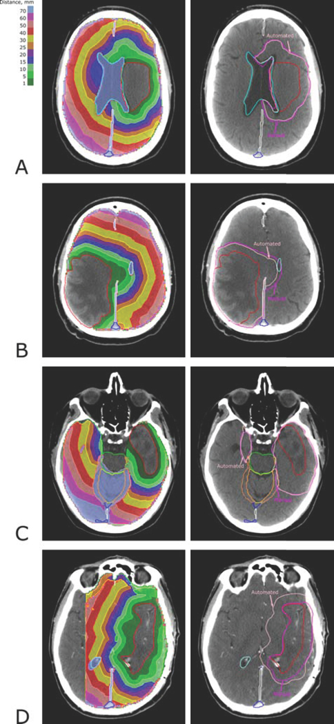
While the CTV itself is ambiguous, it is much easier to identify structures that do not belong to the CTV and serve as barriers to the spread of the disease. We segment the known barrier structures using a convolutional neural network (CNN). The CTV is then obtained by starting from the manually delineated gross tumor volume (GTV) and expanding it while taking into account the barrier structures. Mathematically, we define the CTV as an iso-surface in the 3D map of shortest paths of all voxels from the GTV. The shortest paths are found with the Dijkstra algorithm. While the method is generally applicable, we test it on 206 glioma and glioblastoma cases.
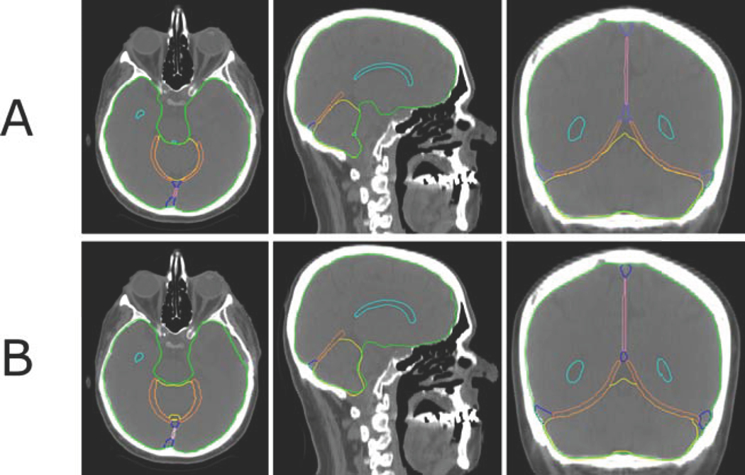
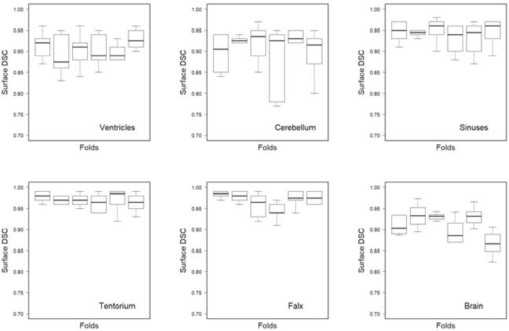
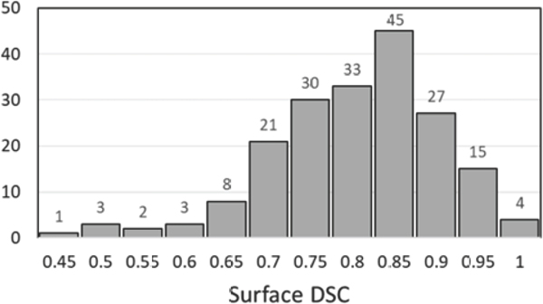
The auto-segmented barrier structures for the brain cases include the ventricles, falx cerebri, tentorium cerebelli, brain sinuses, and the outer surface of the brain. Manual and auto-segmented barrier structures agree with surface Dice Similarity Coefficients (DSC) ranging from 0.91 to 0.97 at 2 mm tolerance. Comparison of manual and automatically delineated CTVs shows a median surface DSC of 0.79.
Barrier structures for CTV definition can be auto-delineated with outstanding precision using a CNN. An algorithm for automated calculation of the CTV by 3D expansion of the GTV while respecting anatomical barriers has been developed. It shows good agreement with manual CTV definition for brain tumors.
临床靶区(CTV)的勾画可以说是治疗计划流程中最薄弱的环节。本研究旨在为临床医生完成这一关键任务提供支持。
虽然 CTV 本身并不明确,但确定不属于 CTV 且作为疾病扩散屏障的结构要容易得多。我们使用卷积神经网络(CNN)对已知的屏障结构进行分割。然后,从手动勾画的大体肿瘤体积(GTV)开始,在考虑到屏障结构的情况下进行扩展,从而获得 CTV。从数学上讲,我们将 CTV 定义为从 GTV 所有体素的最短路径 3D 映射中的等位面。最短路径使用 Dijkstra 算法找到。虽然该方法具有普遍适用性,但我们在 206 例脑胶质瘤和胶质母细胞瘤病例中对其进行了测试。
脑部病例的自动分割的屏障结构包括脑室、大脑镰、小脑幕、脑窦和大脑外表面。手动和自动分割的屏障结构之间的表面 Dice 相似性系数(DSC)在 2mm 容差下的一致性为 0.91 至 0.97。手动和自动勾画的 CTV 之间的比较显示,表面 DSC 的中位数为 0.79。
使用 CNN 可以非常精确地自动勾画 CTV 的屏障结构。已经开发了一种通过 3D 扩展 GTV 并同时尊重解剖学屏障来自动计算 CTV 的算法。它与脑肿瘤的手动 CTV 定义具有良好的一致性。