Perrin Justine, Capitao Marisa, Mougin-Degraef Marie, Guérard François, Faivre-Chauvet Alain, Rbah-Vidal Latifa, Gaschet Joëlle, Guilloux Yannick, Kraeber-Bodéré Françoise, Chérel Michel, Barbet Jacques
CRCINA, INSERM, CNRS, Université d'Angers, Université de Nantes, Nantes, France.
Nuclear Medicine, University Hospital, Nantes, France.
Front Med (Lausanne). 2020 Feb 14;7:34. doi: 10.3389/fmed.2020.00034. eCollection 2020.
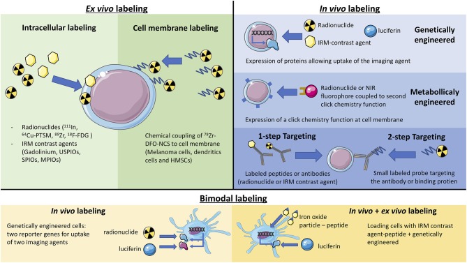
The impressive development of cancer immunotherapy in the last few years originates from a more precise understanding of control mechanisms in the immune system leading to the discovery of new targets and new therapeutic tools. Since different stages of disease progression elicit different local and systemic inflammatory responses, the ability to longitudinally interrogate the migration and expansion of immune cells throughout the whole body will greatly facilitate disease characterization and guide selection of appropriate treatment regiments. While using radiolabeled white blood cells to detect inflammatory lesions has been a classical nuclear medicine technique for years, new non-invasive methods for monitoring the distribution and migration of biologically active cells in living organisms have emerged. They are designed to improve detection sensitivity and allow for a better preservation of cell activity and integrity. These methods include the monitoring of therapeutic cells but also of all cells related to a specific disease or therapeutic approach. Labeling of therapeutic cells for imaging may be performed , with some limitations on sensitivity and duration of observation. Alternatively, cell tracking may be performed by genetically engineering cells or mice so that may be revealed through imaging. In addition, SPECT or PET imaging based on monoclonal antibodies has been used to detect tumors in the human body for years. They may be used to detect and quantify the presence of specific cells within cancer lesions. These methods have been the object of several recent reviews that have concentrated on technical aspects, stressing the differences between direct and indirect labeling. They are briefly described here by distinguishing (labeling cells with paramagnetic, radioactive, or fluorescent tracers) and ( capture of injected radioactive, fluorescent or luminescent tracers, or by using labeled antibodies, ligands, or pre-targeted clickable substrates) imaging methods. This review focuses on cell tracking in specific therapeutic applications, namely cell therapy, and particularly CAR (Chimeric Antigen Receptor) T-cell therapy, which is a fast-growing research field with various therapeutic indications. The potential impact of imaging on the progress of these new therapeutic modalities is discussed.
近年来癌症免疫疗法取得的显著进展源于对免疫系统控制机制更精确的理解,从而发现了新的靶点和新的治疗工具。由于疾病进展的不同阶段会引发不同的局部和全身炎症反应,纵向研究免疫细胞在全身的迁移和扩增能力将极大地促进疾病特征描述,并指导选择合适的治疗方案。虽然使用放射性标记的白细胞检测炎症病变多年来一直是经典的核医学技术,但用于监测活生物体中生物活性细胞分布和迁移的新的非侵入性方法已经出现。它们旨在提高检测灵敏度,并更好地保留细胞活性和完整性。这些方法不仅包括对治疗性细胞的监测,还包括对与特定疾病或治疗方法相关的所有细胞的监测。对治疗性细胞进行成像标记时,在灵敏度和观察持续时间方面存在一些限制。或者,可以通过对细胞或小鼠进行基因工程来进行细胞追踪,以便通过成像揭示细胞情况。此外,基于单克隆抗体的SPECT或PET成像多年来一直用于检测人体肿瘤。它们可用于检测和量化癌症病变内特定细胞的存在。这些方法是最近几篇综述的主题,这些综述集中在技术方面,强调直接标记和间接标记之间的差异。这里通过区分(用顺磁性、放射性或荧光示踪剂标记细胞)和(捕获注入的放射性、荧光或发光示踪剂,或使用标记抗体、配体或预靶向可点击底物)成像方法对它们进行简要描述。本综述重点关注特定治疗应用中的细胞追踪,即细胞治疗,特别是嵌合抗原受体(CAR)T细胞治疗,这是一个快速发展的研究领域,具有各种治疗适应症。本文还讨论了成像对这些新治疗方式进展的潜在影响。