Li Zhipeng, Mu Chunlong, Xu Yixuan, Shen Junshi, Zhu Weiyun
Laboratory of Gastrointestinal Microbiology, Jiangsu Key Laboratory of Gastrointestinal Nutrition and Animal Health, College of Animal Science and Technology, Nanjing Agricultural University, Nanjing, China.
Department of Special Animal Nutrition and Feed Science, Institute of Special Animal and Plant Sciences, Chinese Academy of Agricultural Sciences, Changchun, China.
Front Microbiol. 2020 Feb 21;11:244. doi: 10.3389/fmicb.2020.00244. eCollection 2020.
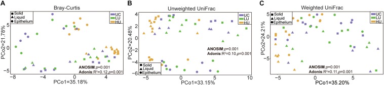
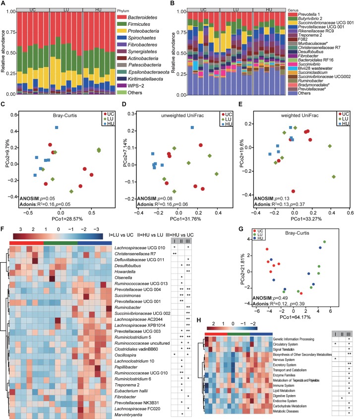
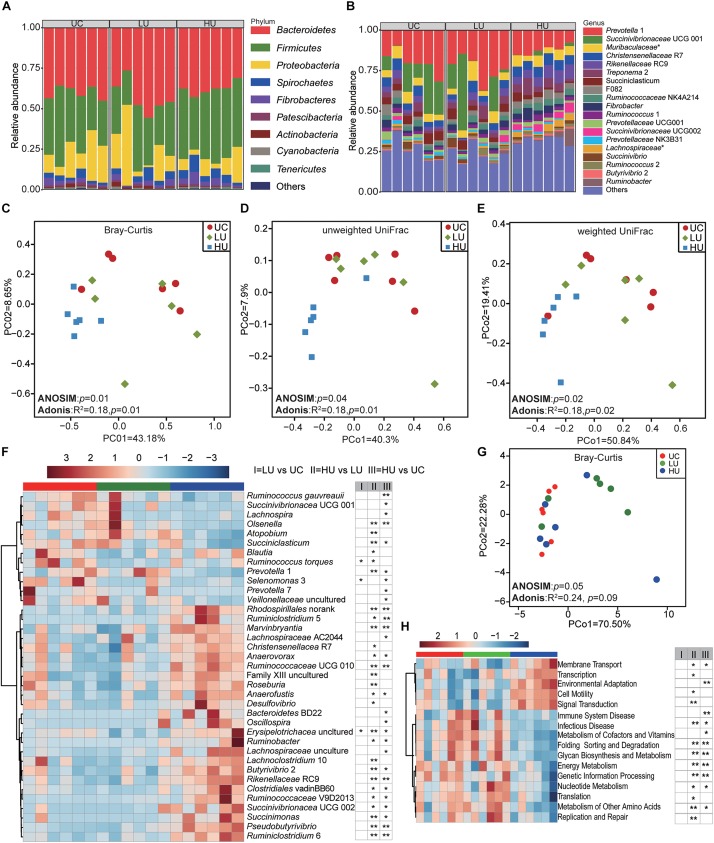
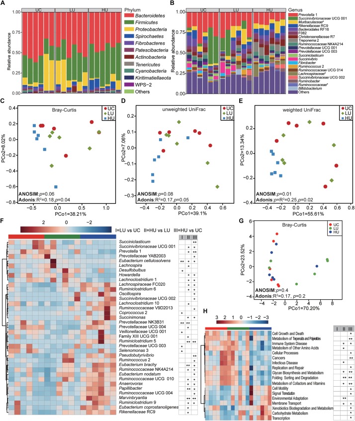
The rumen bacteria in the solid, liquid, and epithelial fractions are distinct and play important roles in the degradation of urea nitrogen. However, the effects of urea on rumen bacteria from the three fractions remain unclear. In this study, 42 Hu lambs were fed a total mixed ration based on concentrate and roughage (55:45, dry matter basis) and randomly assigned to one of three experimental diets: a basal diet with no urea (UC, 0 g/kg), a basal diet supplemented with low urea levels (LU, 10 g/kg DM), and a basal diet supplemented with high urea levels (HU, 30 g/kg DM). After an 11-week feeding trial, six animals from each treatment were harvested. Rumen metabolites levels were measured, and bacteria of the rumen solid, liquid, and epithelial fractions were examined based on 16S rRNA gene sequencing. Under urea supplementation, the concentrations of ammonia and butyrate in the rumen increased, whereas the concentration of propionate decreased. The population of total protozoa was the highest in the LU treatment. 1 was the most abundant genus in all samples. The unclassified , bacteria within the families and , and R7 were abundant in the solid and liquid fractions. 2 and 2 were the abundant bacteria in the epithelial fraction. Principal coordinate analysis showed separation of the solid, liquid and epithelial bacteria regardless of diet, suggesting that rumen fraction had stronger influences on the bacterial community than did urea supplementation. However, the influences on the bacterial community differed among the three fractions. In the solid and liquid fractions, UCG 001 and 1 showed decreased abundance with dietary urea supplementation, whereas the abundance of spp. was increased. spp. and spp. were higher in the epithelial fraction of the UC and LU treatments relative to HU treatment. Comparisons of predictive function in the rumen solid, liquid, and epithelial fractions among the three treatments also revealed differences. Collectively, these results reveal the change of the rumen bacterial community to dietary urea supplementation.
固体、液体和上皮部分中的瘤胃细菌各不相同,在尿素氮的降解中发挥着重要作用。然而,尿素对这三个部分的瘤胃细菌的影响仍不清楚。在本研究中,42只湖羊饲喂基于精料和粗料(干物质基础为55:45)的全混合日粮,并随机分配到三种试验日粮之一:不添加尿素的基础日粮(UC,0 g/kg)、添加低尿素水平的基础日粮(LU,10 g/kg干物质)和添加高尿素水平的基础日粮(HU,30 g/kg干物质)。经过11周的饲养试验,从每个处理中选取6只动物进行屠宰。测定瘤胃代谢物水平,并基于16S rRNA基因测序检查瘤胃固体、液体和上皮部分的细菌。在添加尿素的情况下,瘤胃中氨和丁酸的浓度增加,而丙酸的浓度降低。总原生动物数量在LU处理中最高。1是所有样本中最丰富的属。未分类的、科和科内的细菌以及R7在固体和液体部分中含量丰富。2和2是上皮部分中的优势细菌。主坐标分析表明,无论日粮如何,固体、液体和上皮细菌都有分离,这表明瘤胃部分对细菌群落的影响比添加尿素更强。然而,这三个部分对细菌群落的影响有所不同。在固体和液体部分中,随着日粮中添加尿素,UCG 001和1的丰度降低,而spp.的丰度增加。相对于HU处理,UC和LU处理的上皮部分中spp.和spp.更高。三种处理之间瘤胃固体、液体和上皮部分的预测功能比较也显示出差异。总体而言,这些结果揭示了日粮添加尿素后瘤胃细菌群落的变化。