Kundu Amit Kumar, Fattah Shaikh Anowarul, Wahid Khan A
1Department of Electrical and Electronic EngineeringBangladesh University of Engineering and TechnologyDhaka1205Bangladesh.
2Department of Electrical and Computer EngineeringUniversity of SaskatchewanSaskatoonSKS7N 5A9Canada.
IEEE J Transl Eng Health Med. 2020 Jan 17;8:3300111. doi: 10.1109/JTEHM.2020.2964666. eCollection 2020.
Computer-aided disease detection schemes from wireless capsule endoscopy (WCE) videos have received great attention by the researchers for reducing physicians' burden due to the time-consuming and risky manual review process. While single disease classification schemes are greatly dealt by the researchers in the past, developing a unified scheme which is capable of detecting multiple gastrointestinal (GI) diseases is very challenging due to the highly irregular behavior of diseased images in terms of color patterns.
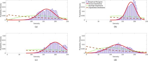
In this paper, a computer-aided method is developed to detect multiple GI diseases from WCE videos utilizing linear discriminant analysis (LDA) based region of interest (ROI) separation scheme followed by a probabilistic model fitting approach. Commonly in training phase, as pixel-labeled images are available in small number, only the image-level annotations are used for detecting diseases in WCE images, whereas pixel-level knowledge, although a major source for learning the disease characteristics, is left unused. In view of learning the characteristic disease patterns from pixel-labeled images, a set of LDA models are trained which are later used to extract the salient ROI from WCE images both in training and testing stages. The intensity patterns of ROI are then modeled by a suitable probability distribution and the fitted parameters of the distribution are utilized as features in a supervised cascaded classification scheme.
For the purpose of validation of the proposed multi-disease detection scheme, a set of pixel-labeled images of bleeding, ulcer and tumor are used to extract the LDA models and then, a large WCE dataset is used for training and testing. A high level of accuracy is achieved even with a small number of pixel-labeled images.
Therefore, the proposed scheme is expected to help physicians in reviewing a large number of WCE images to diagnose different GI diseases.
无线胶囊内窥镜(WCE)视频的计算机辅助疾病检测方案因可减轻医生因耗时且有风险的人工检查过程而产生的负担,受到了研究人员的广泛关注。虽然过去研究人员对单一疾病分类方案进行了大量研究,但由于患病图像在颜色模式方面行为高度不规则,开发一种能够检测多种胃肠道(GI)疾病的统一方案极具挑战性。
本文开发了一种计算机辅助方法,利用基于线性判别分析(LDA)的感兴趣区域(ROI)分离方案,随后采用概率模型拟合方法,从WCE视频中检测多种GI疾病。通常在训练阶段,由于像素标记图像数量较少,仅使用图像级注释来检测WCE图像中的疾病,而像素级知识虽然是学习疾病特征的主要来源,但却未被利用。鉴于从像素标记图像中学习特征性疾病模式,训练了一组LDA模型,随后在训练和测试阶段用于从WCE图像中提取显著ROI。然后,通过合适的概率分布对ROI的强度模式进行建模,并将分布的拟合参数用作监督级联分类方案中的特征。
为了验证所提出的多疾病检测方案,使用一组出血、溃疡和肿瘤的像素标记图像来提取LDA模型,然后,使用一个大型WCE数据集进行训练和测试。即使使用少量像素标记图像也能实现较高的准确率。
因此,预计所提出的方案将有助于医生查看大量WCE图像以诊断不同的GI疾病。