Biemmi Vanessa, Milano Giuseppina, Ciullo Alessandra, Cervio Elisabetta, Burrello Jacopo, Dei Cas Michele, Paroni Rita, Tallone Tiziano, Moccetti Tiziano, Pedrazzini Giovanni, Longnus Sarah, Vassalli Giuseppe, Barile Lucio
Laboratory for Cardiovascular Theranostics, Cardiocentro Ticino Foundation, Lugano, Switzerland.
Faculty of Biomedical Sciences, Università della Svizzera Italiana, Lugano, Switzerland.
Theranostics. 2020 Feb 3;10(6):2773-2790. doi: 10.7150/thno.39072. eCollection 2020.
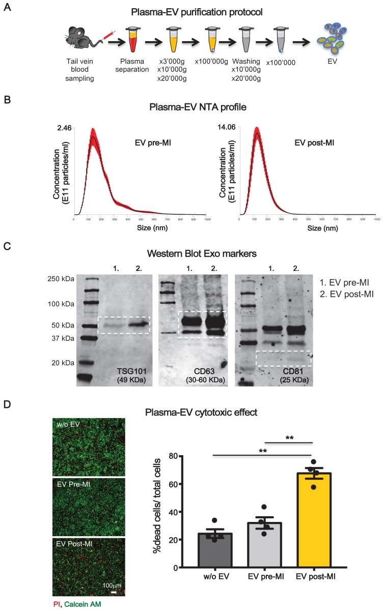
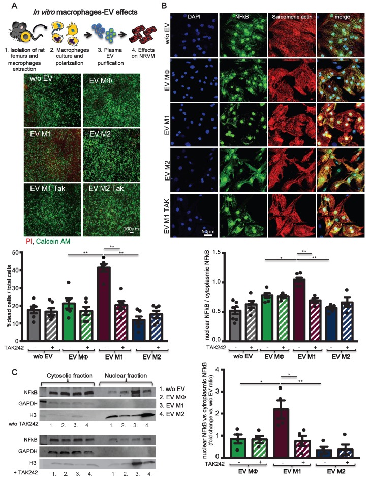
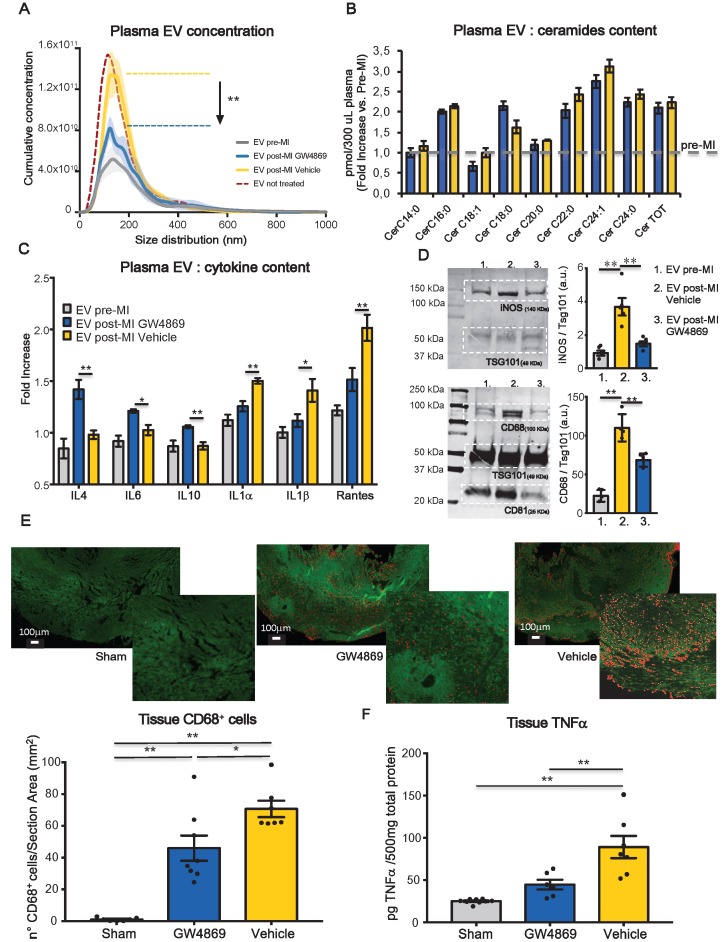
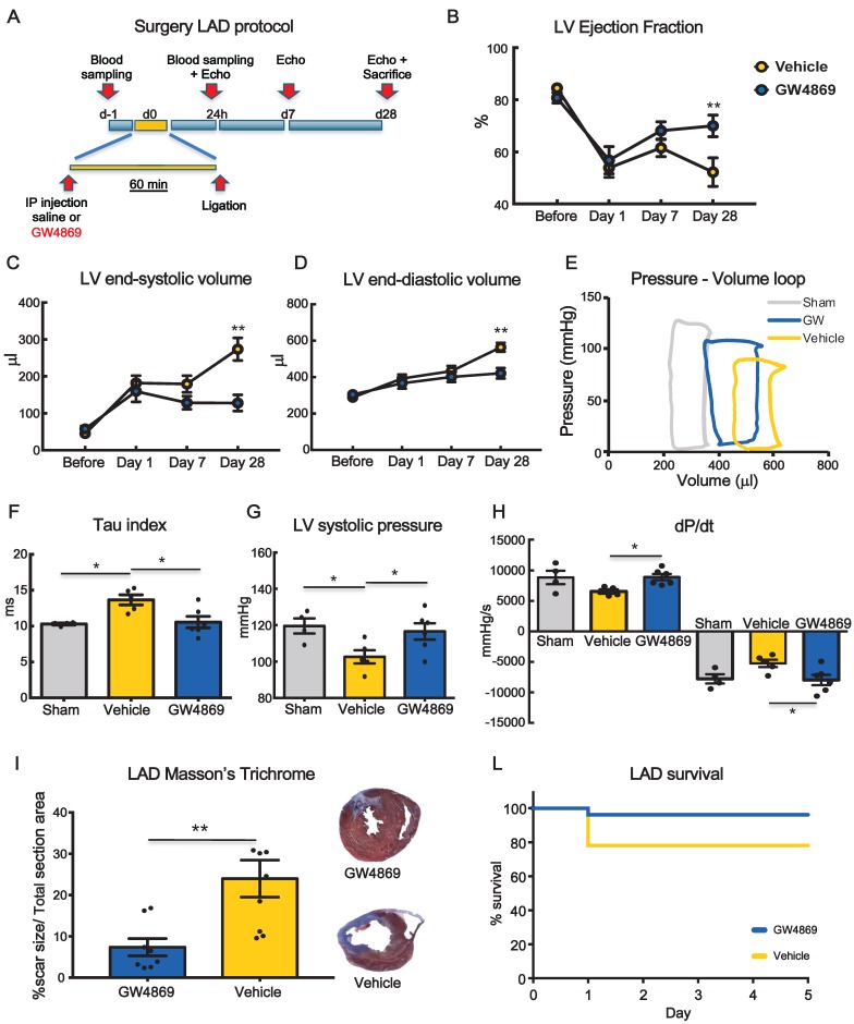
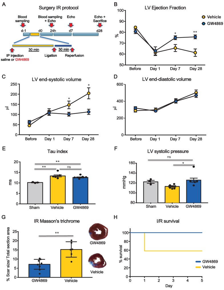
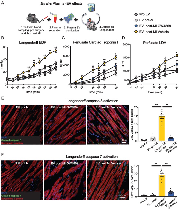
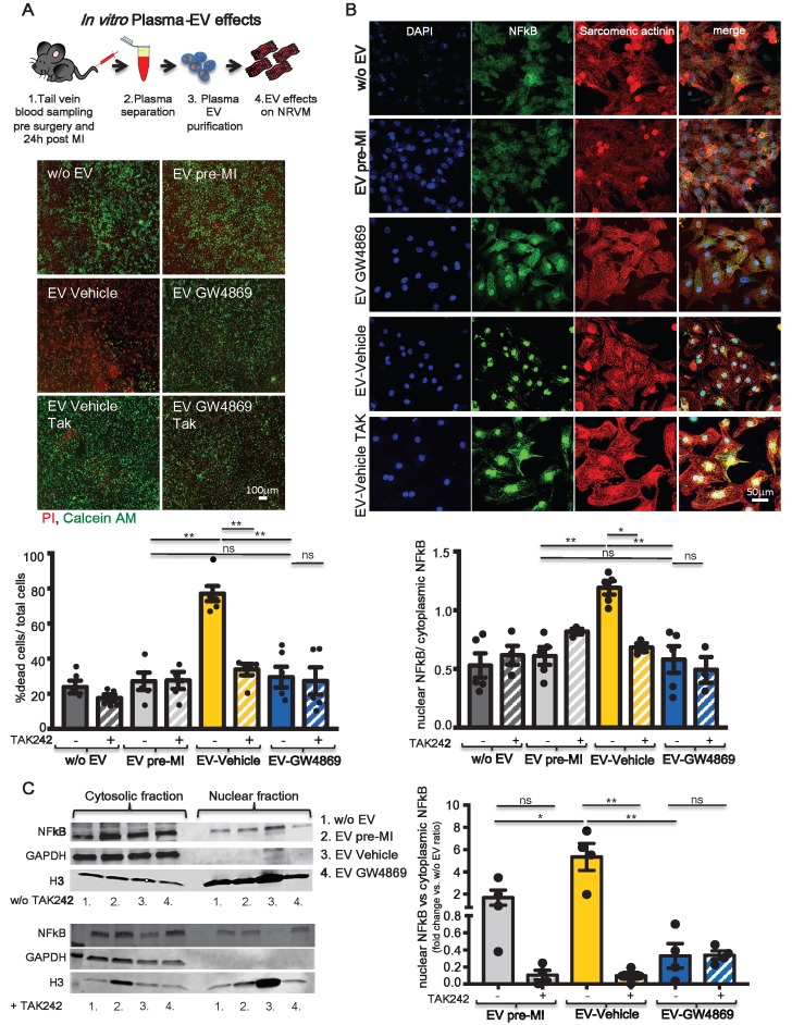
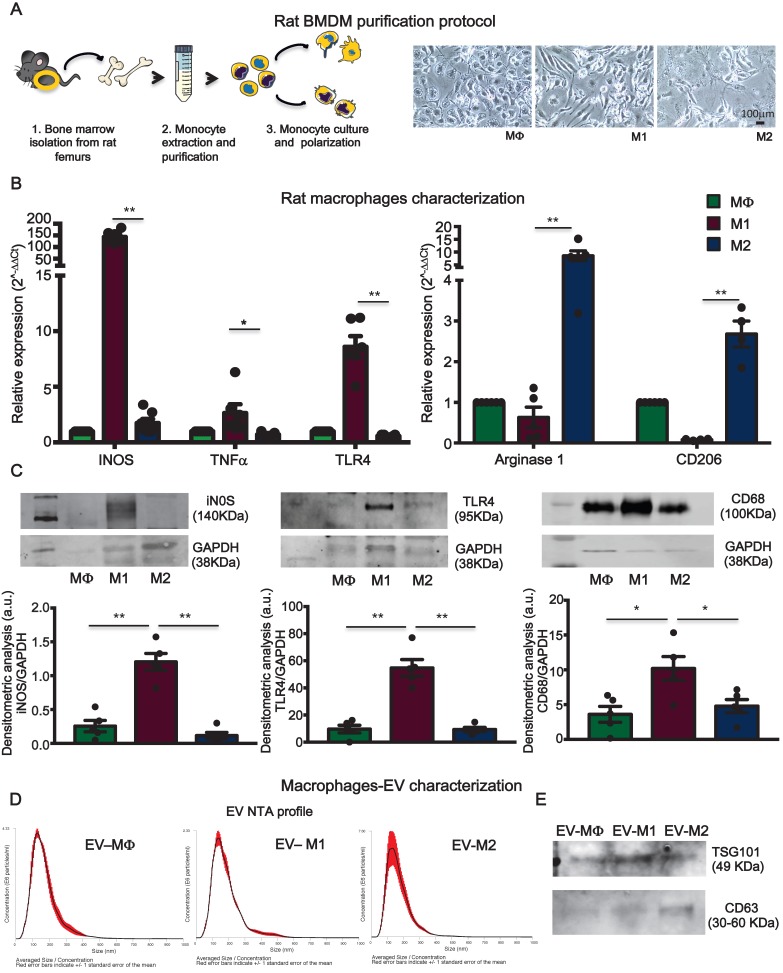
: After myocardial infarction, necrotic cardiomyocytes release damage-associated proteins that stimulate innate immune pathways and macrophage tissue infiltration, which drives inflammation and myocardial remodeling. Circulating inflammatory extracellular vesicles play a crucial role in the acute and chronic phases of ischemia, in terms of inflammatory progression. In this study, we hypothesize that the paracrine effect mediated by these vesicles induces direct cytotoxicity in cardiomyocytes. Thus, we examined whether reducing the generation of inflammatory vesicles within the first few hours after the ischemic event ameliorates cardiac outcome at short and long time points. : Myocardial infarction was induced in rats that were previously injected intraperitoneally with a chemical inhibitor of extracellular-vesicle biogenesis. Heart global function was assessed by echocardiography performed at 7, 14 and 28 days after MI. Cardiac outcome was also evaluated by hemodynamic analysis at sacrifice. Cytotoxic effects of circulating EV were evaluated in a Langendorff, system by measuring the level of cardiac troponin I (cTnI) in the perfusate. Mechanisms undergoing cytotoxic effects of EV derived from pro-inflammatory macrophages (M1) were studied in primary rat neonatal cardiomyocytes. : Inflammatory response following myocardial infarction dramatically increased the number of circulating extracellular vesicles carrying alarmins such as IL-1α, IL-1β and Rantes. Reducing the boost in inflammatory vesicles during the acute phase of ischemia resulted in preserved left ventricular ejection fraction . Hemodynamic analysis confirmed functional recovery by displaying higher velocity of left ventricular relaxation and improved contractility. When added to the perfusate of isolated hearts, post-infarction circulating vesicles induced significantly more cell death in adult cardiomyocytes, as assessed by cTnI release, comparing to circulating vesicles isolated from healthy (non-infarcted) rats. inflammatory extracellular vesicles induce cell death by driving nuclear translocation of NF-κB into nuclei of cardiomyocytes. : Our data suggest that targeting circulating extracellular vesicles during the acute phase of myocardial infarction may offer an effective therapeutic approach to preserve function of ischemic heart.
心肌梗死后,坏死的心肌细胞释放损伤相关蛋白,刺激固有免疫途径和巨噬细胞组织浸润,从而引发炎症和心肌重塑。就炎症进展而言,循环炎性细胞外囊泡在缺血的急性期和慢性期起着关键作用。在本研究中,我们假设这些囊泡介导的旁分泌效应会在心肌细胞中诱导直接细胞毒性。因此,我们研究了在缺血事件发生后的最初几个小时内减少炎性囊泡的产生是否能在短期和长期改善心脏结局。:对先前腹腔注射细胞外囊泡生物合成化学抑制剂的大鼠诱导心肌梗死。在心肌梗死后7天、14天和28天通过超声心动图评估心脏整体功能。在处死时通过血流动力学分析评估心脏结局。通过测量灌注液中心肌肌钙蛋白I(cTnI)的水平,在Langendorff系统中评估循环细胞外囊泡的细胞毒性作用。在原代大鼠新生心肌细胞中研究源自促炎性巨噬细胞(M1)的细胞外囊泡细胞毒性作用的机制。:心肌梗死后的炎症反应显著增加了携带警报素如IL-1α、IL-1β和RANTES的循环细胞外囊泡的数量。在缺血急性期减少炎性囊泡的增加可使左心室射血分数得以保留。血流动力学分析通过显示更高的左心室舒张速度和改善的收缩性证实了功能恢复。与从健康(未梗死)大鼠分离的循环囊泡相比,当将梗死后期循环囊泡添加到离体心脏的灌注液中时,通过cTnI释放评估,其在成年心肌细胞中诱导的细胞死亡明显更多。炎性细胞外囊泡通过促使NF-κB核转位进入心肌细胞核而诱导细胞死亡。:我们的数据表明,在心肌梗死急性期靶向循环细胞外囊泡可能提供一种有效的治疗方法来保留缺血心脏的功能。