Department of Oral and Maxillofacial Surgery and Central Laboratory, School and Hospital of Stomatology, Peking University, No. 22 Zhong-Guan-Cun South Road, Hai-Dian District, Beijing, 100081, China.
Laboratory of Biomaterials and Regenerative Medicine, Academy for Advanced Interdisciplinary Studies, Peking University, Beijing, 100871, China.
Stem Cell Res Ther. 2020 Mar 20;11(1):127. doi: 10.1186/s13287-020-01628-4.
Organ replacement regenerative therapy based on human adult stem cells may be effective for salivary gland hypofunction. However, the generated tissues are immature because the signaling factors that induce the differentiation of human salivary gland stem cells into salivary glands are unknown.
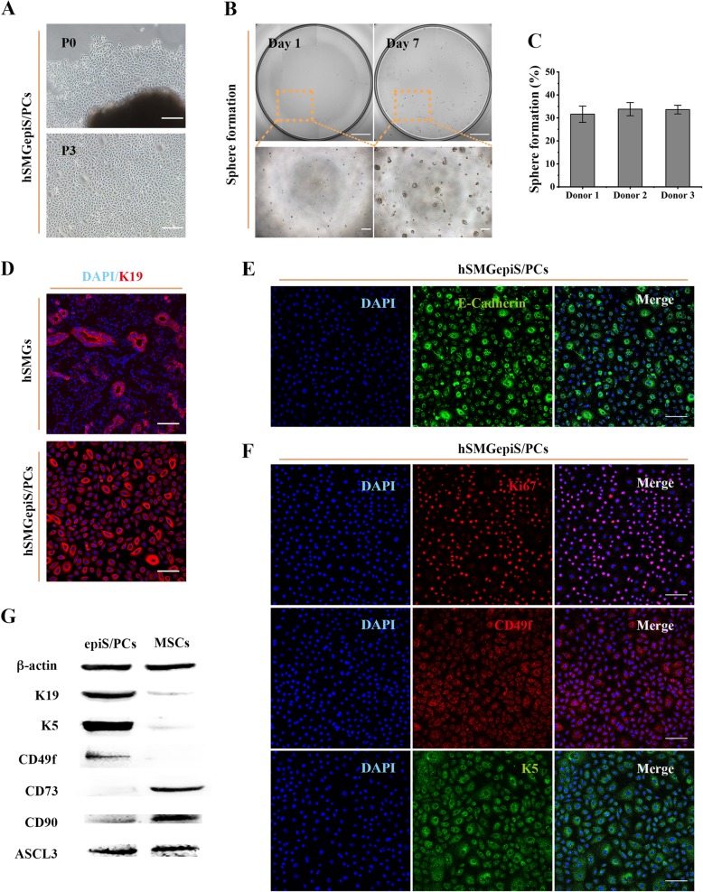
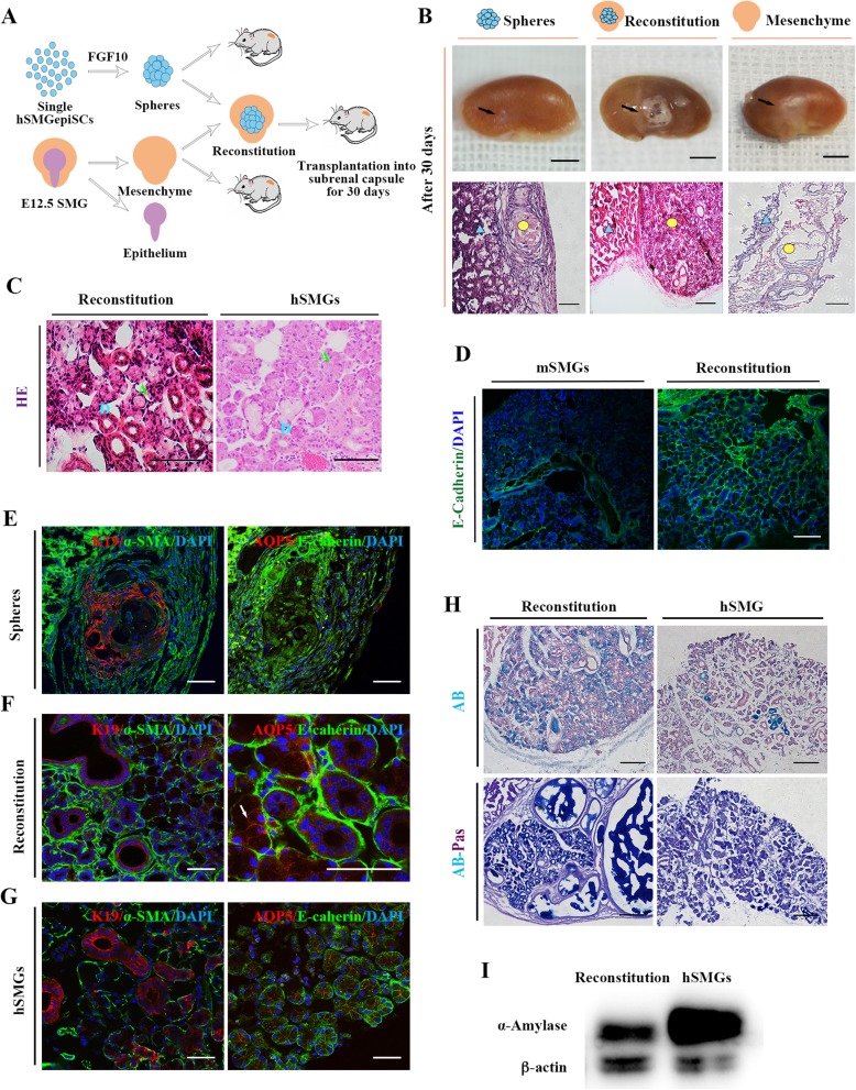
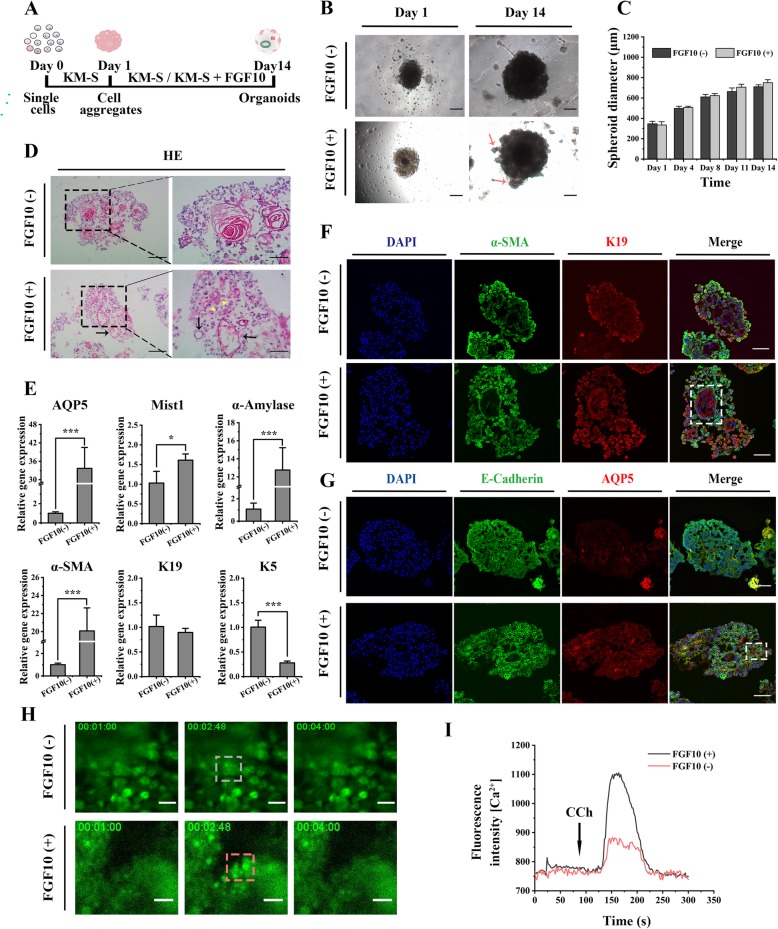
Isolated human submandibular gland stem/progenitor cells (hSMGepiS/PCs) were characterized and three-dimensionally (3D) cultured to generate organoids and further induced by fibroblast growth factor 10 (FGF10) in vitro. The induced spheres alone or in combination with embryonic day 12.5 (E12.5) mouse salivary gland mesenchyme were transplanted into the renal capsules of nude mice to assess their development in vivo. Immunofluorescence, quantitative reverse transcriptase-polymerase chain reaction, calcium release analysis, western blotting, hematoxylin-eosin staining, Alcian blue-periodic acid-Schiff staining, and Masson's trichrome staining were performed to assess the structure and function of generated tissues in vitro and in vivo.
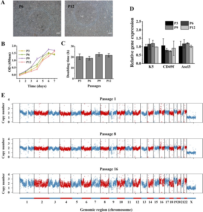
The isolated hSMGepiS/PCs could be long-term cultured with a stable genome. The organoids treated with FGF10 [FGF10 (+) group] exhibited higher expression of salivary gland-specific markers; showed spatial arrangement of AQP5, K19, and SMA cells; and were more sensitive to the stimulation by neurotransmitters than untreated organoids [FGF10 (-) group]. After heterotopic transplantation, the induced cell spheres combined with mouse embryonic salivary gland mesenchyme showed characteristics of mature salivary glands, including a natural morphology and saliva secretion.
FGF10 promoted the development of the hSMGepiS/PC-derived salivary gland organoids by the expression of differentiation markers, structure formation, and response to neurotransmitters in vitro. Moreover, the hSMGepiS/PCs responded to the niche in mouse embryonic mesenchyme and further differentiated into salivary gland tissues with mature characteristics. Our study provides a foundation for the regenerative therapy of salivary gland diseases.
基于人成体干细胞的器官替代再生疗法可能对唾液腺功能低下有效。然而,由于诱导人唾液腺干细胞分化为唾液腺的信号因子尚不清楚,因此产生的组织不成熟。
分离人下颌下腺干细胞/祖细胞(hSMGepiS/PC)并进行三维(3D)培养以生成类器官,然后在体外进一步用成纤维细胞生长因子 10(FGF10)诱导。单独诱导的球体或与胚胎第 12.5 天(E12.5)的小鼠唾液腺间充质一起移植到裸鼠肾囊中,以评估其体内发育情况。免疫荧光、实时定量逆转录-聚合酶链反应、钙释放分析、Western blot、苏木精-伊红染色、阿辛蓝-过碘酸-希夫染色和马松三色染色用于评估体外和体内生成组织的结构和功能。
分离的 hSMGepiS/PC 可长期稳定培养。用 FGF10 处理的类器官[FGF10(+)组]表现出更高的唾液腺特异性标志物表达;显示 AQP5、K19 和 SMA 细胞的空间排列;并且对神经递质的刺激比未经处理的类器官[FGF10(-)组]更敏感。异位移植后,诱导的细胞球与小鼠胚胎唾液腺间充质结合显示出成熟唾液腺的特征,包括自然形态和唾液分泌。
FGF10 在体外通过分化标志物的表达、结构形成和对神经递质的反应促进了 hSMGepiS/PC 衍生的唾液腺类器官的发育。此外,hSMGepiS/PC 对小鼠胚胎间充质的小生境有反应,并进一步分化为具有成熟特征的唾液腺组织。我们的研究为唾液腺疾病的再生治疗提供了基础。