• 文献检索
  • 文档翻译
  • 深度研究
  • 学术资讯
  • Suppr Zotero 插件Zotero 插件
  • 邀请有礼
  • 套餐&价格
  • 历史记录
应用&插件
Suppr Zotero 插件Zotero 插件浏览器插件Mac 客户端Windows 客户端微信小程序
定价
高级版会员购买积分包购买API积分包
服务
文献检索文档翻译深度研究API 文档MCP 服务
关于我们
关于 Suppr公司介绍联系我们用户协议隐私条款
关注我们

Suppr 超能文献

核心技术专利:CN118964589B侵权必究
粤ICP备2023148730 号-1Suppr @ 2026

文献检索

告别复杂PubMed语法,用中文像聊天一样搜索,搜遍4000万医学文献。AI智能推荐,让科研检索更轻松。

立即免费搜索

文件翻译

保留排版,准确专业,支持PDF/Word/PPT等文件格式,支持 12+语言互译。

免费翻译文档

深度研究

AI帮你快速写综述,25分钟生成高质量综述,智能提取关键信息,辅助科研写作。

立即免费体验

尼曼-匹克病 C 型的视网膜轴突变性。

Retinal axonal degeneration in Niemann-Pick type C disease.

机构信息

Institute of Clinical Neuroimmunology, Ludwig-Maximilians University, Marchioninistr. 15, 81377, Munich, Germany.

Department of Neurology, Ludwig-Maximilians University München, Marchioninistr. 15, 81377, Munich, Germany.

出版信息

J Neurol. 2020 Jul;267(7):2070-2082. doi: 10.1007/s00415-020-09796-2. Epub 2020 Mar 28.

DOI:10.1007/s00415-020-09796-2
PMID:32222928
原文链接:https://pmc.ncbi.nlm.nih.gov/articles/PMC7320959/
Abstract

OBJECTIVE

Niemann-Pick disease type C1 (NPC1) is a rare autosomal-recessive lysosomal storage disorder presenting with a broad clinical spectrum ranging from a severe infantile-onset neurovisceral disorder to late-onset neurodegenerative disease. Optical coherence tomography (OCT) is established to detect retinal degeneration in vivo. We examined NPC1-patients (NPC1-P), clinically asymptomatic NPC1-mutation carriers (NPC1-MC), and healthy controls (HC) to (1) identify retinal degeneration in NPC1-disease and (2) to investigate possible subclinical retinal degeneration in NPC1-MC.

METHODS

Fourteen NPC1-P, 17 NPC1-MC, and 31 age-matched HC were examined using spectral-domain OCT. Neurological examinations, clinical scales [modified Disability Rating Scale (mDRS); Scale for the Rating and Assessment of Ataxia (SARA); Spinocerebellar Ataxia Functional Index (SCAFI)], and video-oculography (VOG) were correlated with OCT data.

RESULTS

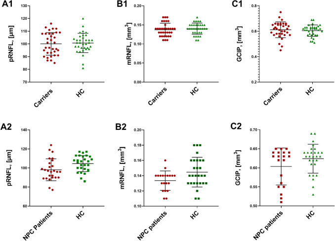
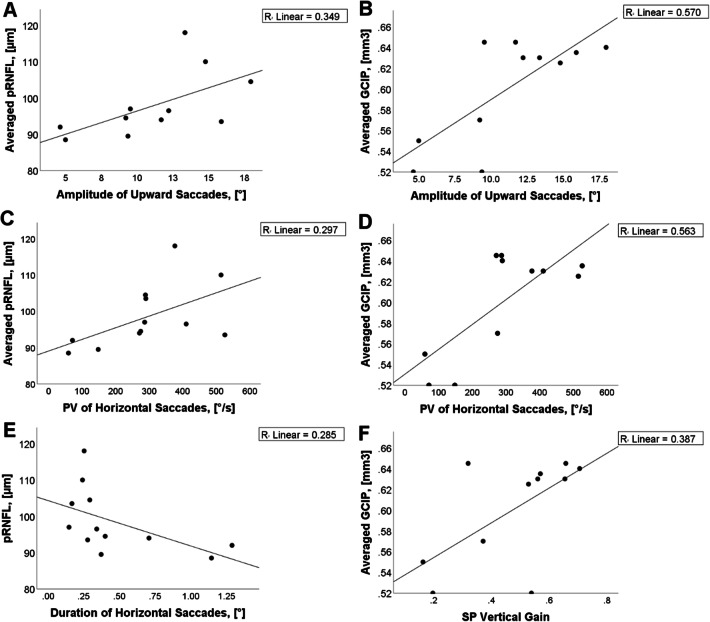
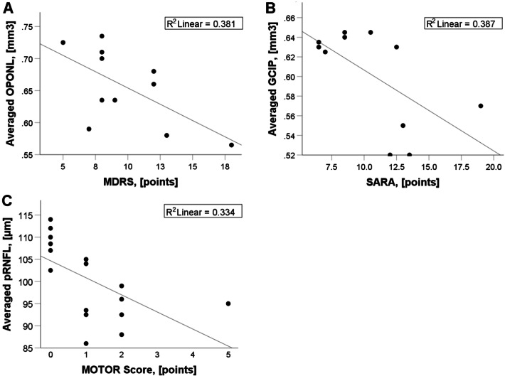
Macular retinal nerve fiber layer and volumes of combined ganglion cell and inner plexiform layer were significantly lower in NPC1-P compared to HC [mRNFL (µm):0.13 ± 0.01 vs. 0.14 ± 0.02; p = 0.01; GCIPL (mm):0.60 ± 0.05 vs. 0.62 ± 0.04; p = 0.04]. No significant differences were found in NPC1-MC in comparison to HC. In NPC1-P, the amplitude of upward vertical saccades showed positive associations with peripapillary RNFL (ρ = 0.645; p < 0.05), and thinned GCIP (ρ = 0.609; p < 0.05), but not in NPC1-MC. In NPC1-P correlations between combined outer plexiform layer and outer nuclear layer (OPONL) with mDRS (r = - 0.617; p < 0.05) and GCIP with SARA (r = - 0.622; p < 0.05) were observed. Furthermore, in NPC1-MC, motor scores were negatively associated with pRNFL (ρ = - 0.677; p < 0.01).

CONCLUSIONS

Using OCT, we showed retinal degeneration in NPC1-P and significant correlation between retinal neuroaxonal degeneration with clinical measurements. We observed a non-significant trend of retinal degeneration in NPC1-MC correlating with subclinical motor abnormalities. Based on these preliminary data, OCT may be an important marker of neurodegeneration in NPC1-disease after onset of clinical symptoms.

摘要

目的

尼曼-皮克病 C1 型(NPC1)是一种罕见的常染色体隐性溶酶体贮积症,具有广泛的临床谱,从严重的婴儿发病神经内脏障碍到迟发性神经退行性疾病。光学相干断层扫描(OCT)被用于检测体内的视网膜变性。我们检查了 NPC1 患者(NPC1-P)、临床无症状 NPC1 突变携带者(NPC1-MC)和健康对照者(HC),以(1)识别 NPC1 疾病中的视网膜变性,(2)研究 NPC1-MC 中可能存在的亚临床视网膜变性。

方法

使用谱域 OCT 检查了 14 名 NPC1-P、17 名 NPC1-MC 和 31 名年龄匹配的 HC。对神经学检查、临床量表[改良残疾评分量表(mDRS);共济失调评定量表(SARA);脊髓小脑共济失调功能指数(SCAFI)]和视频眼动图(VOG)与 OCT 数据进行了相关性分析。

结果

与 HC 相比,NPC1-P 的黄斑视网膜神经纤维层和节细胞和内丛状层的容积明显降低[mRNFL(µm):0.13±0.01 对 0.14±0.02;p=0.01;GCIPL(mm):0.60±0.05 对 0.62±0.04;p=0.04]。与 HC 相比,NPC1-MC 无明显差异。在 NPC1-P 中,向上垂直扫视的振幅与视盘周围 RNFL(ρ=0.645;p<0.05)和变薄的 GCIP(ρ=0.609;p<0.05)呈正相关,但在 NPC1-MC 中则无相关性。在 NPC1-P 中,观察到外丛状层和外核层(OPONL)与 mDRS(r=-0.617;p<0.05)和 GCIP 与 SARA(r=-0.622;p<0.05)之间存在相关性。此外,在 NPC1-MC 中,运动评分与 pRNFL 呈负相关(ρ=-0.677;p<0.01)。

结论

使用 OCT,我们显示 NPC1-P 存在视网膜变性,并且视网膜神经轴突变性与临床测量之间存在显著相关性。我们观察到 NPC1-MC 存在非显著性的视网膜变性趋势,与亚临床运动异常相关。基于这些初步数据,OCT 可能是 NPC1 疾病临床症状出现后神经退行性变的一个重要标志物。

https://cdn.ncbi.nlm.nih.gov/pmc/blobs/a45a/7320959/ba189dbe706c/415_2020_9796_Fig3_HTML.jpg
https://cdn.ncbi.nlm.nih.gov/pmc/blobs/a45a/7320959/f03450390716/415_2020_9796_Fig1_HTML.jpg
https://cdn.ncbi.nlm.nih.gov/pmc/blobs/a45a/7320959/57341ca34941/415_2020_9796_Fig2_HTML.jpg
https://cdn.ncbi.nlm.nih.gov/pmc/blobs/a45a/7320959/ba189dbe706c/415_2020_9796_Fig3_HTML.jpg
https://cdn.ncbi.nlm.nih.gov/pmc/blobs/a45a/7320959/f03450390716/415_2020_9796_Fig1_HTML.jpg
https://cdn.ncbi.nlm.nih.gov/pmc/blobs/a45a/7320959/57341ca34941/415_2020_9796_Fig2_HTML.jpg
https://cdn.ncbi.nlm.nih.gov/pmc/blobs/a45a/7320959/ba189dbe706c/415_2020_9796_Fig3_HTML.jpg

相似文献

1
Retinal axonal degeneration in Niemann-Pick type C disease.尼曼-匹克病 C 型的视网膜轴突变性。
J Neurol. 2020 Jul;267(7):2070-2082. doi: 10.1007/s00415-020-09796-2. Epub 2020 Mar 28.
2
Vertical saccadic palsy and foveal retinal thinning in Niemann-Pick disease type C.尼曼-匹克病 C 型患者的垂直性眼球跳动麻痹和中心凹视网膜变薄。
PLoS One. 2021 Jun 4;16(6):e0252825. doi: 10.1371/journal.pone.0252825. eCollection 2021.
3
Retinal imaging and axonal degeneration in later onset multiple sclerosis.迟发性多发性硬化症中的视网膜成像与轴突退变
J Neurol Sci. 2016 Nov 15;370:1-6. doi: 10.1016/j.jns.2016.09.009. Epub 2016 Sep 8.
4
Thickness of macular inner retinal layers and peripapillary retinal nerve fibre layer in neuromyelitis optica spectrum optic neuritis and isolated optic neuritis with one episode.视神经脊髓炎谱系疾病相关性视神经炎及单次发作的孤立性视神经炎中黄斑区视网膜内层及视乳头周围视网膜神经纤维层的厚度
Acta Ophthalmol. 2017 Sep;95(6):583-590. doi: 10.1111/aos.13257. Epub 2016 Oct 24.
5
Ganglion cell-inner plexiform layer and retinal nerve fibre layer changes within the macula in retinitis pigmentosa: a spectral domain optical coherence tomography study.色素性视网膜炎黄斑部神经节细胞-内丛状层和视网膜神经纤维层的改变:频域光学相干断层扫描研究。
Acta Ophthalmol. 2018 Mar;96(2):e180-e188. doi: 10.1111/aos.13577. Epub 2017 Nov 2.
6
Topographic localization of macular retinal ganglion cell loss associated with localized peripapillary retinal nerve fiber layer defect.黄斑视网膜神经节细胞丢失与局限性视盘周围视网膜神经纤维层缺损的定位关系。
Invest Ophthalmol Vis Sci. 2014 May 6;55(6):3501-8. doi: 10.1167/iovs.14-13925.
7
Clinical, ocular motor, and imaging profile of Niemann-Pick type C heterozygosity.尼曼-皮克 C 型杂合子的临床、眼动和影像学特征。
Neurology. 2020 Apr 21;94(16):e1702-e1715. doi: 10.1212/WNL.0000000000009290. Epub 2020 Mar 31.
8
Segmented inner plexiform layer thickness as a potential biomarker to evaluate open-angle glaucoma: Dendritic degeneration of retinal ganglion cell.分段内网状层厚度作为评估开角型青光眼的潜在生物标志物:视网膜神经节细胞的树突状变性
PLoS One. 2017 Aug 3;12(8):e0182404. doi: 10.1371/journal.pone.0182404. eCollection 2017.
9
Localization of Niemann-Pick C1 protein in astrocytes: implications for neuronal degeneration in Niemann- Pick type C disease.尼曼-皮克C1蛋白在星形胶质细胞中的定位:对尼曼-皮克C型疾病中神经元变性的影响。
Proc Natl Acad Sci U S A. 1999 Feb 16;96(4):1657-62. doi: 10.1073/pnas.96.4.1657.
10
Repeatability of peripapillary retinal nerve fiber layer and inner retinal thickness among two spectral domain optical coherence tomography devices.两种光谱域光学相干断层扫描设备测量视乳头周围视网膜神经纤维层及视网膜内层厚度的可重复性
Invest Ophthalmol Vis Sci. 2014 Sep 16;55(10):6536-46. doi: 10.1167/iovs.14-15072.

引用本文的文献

1
Best Oculomotor Endpoints for Clinical Trials in Hereditary Ataxias: A Systematic Review and Consensus by the Ataxia Global Initiative Working Group on Digital‑Motor Biomarkers.遗传性共济失调临床试验的最佳动眼神经终点:共济失调全球倡议数字运动生物标志物工作组的系统评价与共识
Cerebellum. 2025 Aug 13;24(5):141. doi: 10.1007/s12311-025-01894-z.
2
Differential gene expression patterns in Niemann-Pick Type C and Tay-Sachs diseases: Implications for neurodegenerative mechanisms.尼曼-皮克病C型和泰-萨克斯病中的差异基因表达模式:对神经退行性机制的启示
PLoS One. 2025 Mar 19;20(3):e0319401. doi: 10.1371/journal.pone.0319401. eCollection 2025.
3

本文引用的文献

1
Seeing function in structure: "incidental" eye findings on OCT in a patient with multiple sclerosis.从结构中观察功能:多发性硬化症患者 OCT 上的“偶然”眼部发现。
Mult Scler Relat Disord. 2019 Jun;31:72-73. doi: 10.1016/j.msard.2019.03.019. Epub 2019 Mar 28.
2
CSI: Multiple sclerosis. Tracing optic nerve involvement by standardized optical coherence tomography.犯罪现场调查:多发性硬化症。通过标准化光学相干断层扫描追踪视神经受累情况。
Ann Neurol. 2019 May;85(5):615-617. doi: 10.1002/ana.25471. Epub 2019 Apr 10.
3
Optical coherence tomography: a window to the optic nerve in clinically isolated syndrome.
Peripapillary Retinal Nerve Fiber Layer (pRNFL) Thickness - A Novel Biomarker of Neurodegeneration in Late-Infantile CLN2 Disease.
视乳头周围视网膜神经纤维层(pRNFL)厚度——晚发性婴儿型CLN2病神经退行性变的一种新型生物标志物。
Eye Brain. 2024 Nov 13;16:101-113. doi: 10.2147/EB.S473408. eCollection 2024.
4
[Retinal OCT biomarkers and neurodegenerative diseases of the central nervous system beyond Alzheimer's disease].[视网膜光学相干断层扫描生物标志物与阿尔茨海默病以外的中枢神经系统神经退行性疾病]
Ophthalmologie. 2024 Feb;121(2):93-104. doi: 10.1007/s00347-023-01974-7. Epub 2024 Jan 23.
5
Sigma-2 Receptors-From Basic Biology to Therapeutic Target: A Focus on Age-Related Degenerative Diseases.Sigma-2 受体——从基础生物学到治疗靶点:关注与年龄相关的退行性疾病。
Int J Mol Sci. 2023 Mar 26;24(7):6251. doi: 10.3390/ijms24076251.
6
Quantitative Oculomotor Assessment in Hereditary Ataxia: Discriminatory Power, Correlation with Severity Measures, and Recommended Parameters for Specific Genotypes.遗传性共济失调的定量眼动评估:鉴别能力、与严重度测量的相关性,以及特定基因型的推荐参数。
Cerebellum. 2024 Feb;23(1):121-135. doi: 10.1007/s12311-023-01514-8. Epub 2023 Jan 14.
7
Dynamic lipid turnover in photoreceptors and retinal pigment epithelium throughout life.感光细胞和视网膜色素上皮细胞中动态脂质周转在整个生命过程中。
Prog Retin Eye Res. 2022 Jul;89:101037. doi: 10.1016/j.preteyeres.2021.101037. Epub 2021 Dec 29.
8
Optical Coherence Tomography to Assess Neurodegeneration in Phenylalanine Hydroxylase Deficiency.光学相干断层扫描评估苯丙氨酸羟化酶缺乏症中的神经退行性变
Front Neurol. 2021 Dec 10;12:780624. doi: 10.3389/fneur.2021.780624. eCollection 2021.
9
International consensus on clinical severity scale use in evaluating Niemann-Pick disease Type C in paediatric and adult patients: results from a Delphi Study.国际共识:临床严重程度量表在评估儿科和成年尼曼-匹克病 C 型患者中的应用:德尔菲研究结果。
Orphanet J Rare Dis. 2021 Nov 18;16(1):482. doi: 10.1186/s13023-021-02115-6.
10
A cross-sectional, prospective ocular motor study in 72 patients with Niemann-Pick disease type C.72 例尼曼-匹克病 C 型患者的横断面前瞻性眼动研究。
Eur J Neurol. 2021 Sep;28(9):3040-3050. doi: 10.1111/ene.14955. Epub 2021 Jul 12.
光学相干断层扫描:临床孤立综合征视神经的窗口。
Brain. 2019 Apr 1;142(4):903-915. doi: 10.1093/brain/awz038.
4
Association of Retinal Neurodegeneration on Optical Coherence Tomography With Dementia: A Population-Based Study.基于人群的研究:光学相干断层扫描视网膜神经退行性变与痴呆的相关性。
JAMA Neurol. 2018 Oct 1;75(10):1256-1263. doi: 10.1001/jamaneurol.2018.1563.
5
Retinal layer segmentation in multiple sclerosis: a systematic review and meta-analysis.多发性硬化症中的视网膜层分割:系统评价和荟萃分析。
Lancet Neurol. 2017 Oct;16(10):797-812. doi: 10.1016/S1474-4422(17)30278-8. Epub 2017 Sep 12.
6
Repeatability and Reproducibility of Retinal Neuronal and Axonal Measures on Spectral-Domain Optical Coherence Tomography in Patients with Cognitive Impairment.认知障碍患者视网膜神经元和轴突测量在光谱域光学相干断层扫描中的可重复性和再现性
Front Neurol. 2017 Aug 15;8:359. doi: 10.3389/fneur.2017.00359. eCollection 2017.
7
Optical Coherence Tomography Reveals Retinal Neuroaxonal Thinning in Frontotemporal Dementia as in Alzheimer's Disease.光学相干断层扫描显示,额颞叶痴呆患者的视网膜神经轴突变薄,与阿尔茨海默病患者情况相同。
J Alzheimers Dis. 2017;56(3):1101-1107. doi: 10.3233/JAD-160886.
8
Imaging retina to study dementia and stroke.利用影像学检查视网膜以研究痴呆和中风。
Prog Retin Eye Res. 2017 Mar;57:89-107. doi: 10.1016/j.preteyeres.2017.01.001. Epub 2017 Jan 3.
9
Contribution of cholesterol and oxysterols to the pathophysiology of Parkinson's disease.胆固醇和氧化甾醇在帕金森病病理生理学中的作用。
Free Radic Biol Med. 2016 Dec;101:393-400. doi: 10.1016/j.freeradbiomed.2016.10.008. Epub 2016 Nov 9.
10
Role of Niemann-Pick Type C Disease Mutations in Dementia.尼曼-匹克C型病突变在痴呆症中的作用。
J Alzheimers Dis. 2017;55(3):1249-1259. doi: 10.3233/JAD-160214.