• 文献检索
  • 文档翻译
  • 深度研究
  • 学术资讯
  • Suppr Zotero 插件Zotero 插件
  • 邀请有礼
  • 套餐&价格
  • 历史记录
应用&插件
Suppr Zotero 插件Zotero 插件浏览器插件Mac 客户端Windows 客户端微信小程序
定价
高级版会员购买积分包购买API积分包
服务
文献检索文档翻译深度研究API 文档MCP 服务
关于我们
关于 Suppr公司介绍联系我们用户协议隐私条款
关注我们

Suppr 超能文献

核心技术专利:CN118964589B侵权必究
粤ICP备2023148730 号-1Suppr @ 2026

文献检索

告别复杂PubMed语法,用中文像聊天一样搜索,搜遍4000万医学文献。AI智能推荐,让科研检索更轻松。

立即免费搜索

文件翻译

保留排版,准确专业,支持PDF/Word/PPT等文件格式,支持 12+语言互译。

免费翻译文档

深度研究

AI帮你快速写综述,25分钟生成高质量综述,智能提取关键信息,辅助科研写作。

立即免费体验

认知障碍患者视网膜神经元和轴突测量在光谱域光学相干断层扫描中的可重复性和再现性

Repeatability and Reproducibility of Retinal Neuronal and Axonal Measures on Spectral-Domain Optical Coherence Tomography in Patients with Cognitive Impairment.

作者信息

Loh Edwin Hong-Teck, Ong Yi-Ting, Venketasubramanian Narayanaswamy, Hilal Saima, Thet Naing, Wong Tien Yin, Chen Christopher P L, Cheung Carol Yim-Lui

机构信息

Singapore Eye Research Institute, Singapore National Eye Centre, Singapore, Singapore.

Duke-NUS Medical School, National University of Singapore, Singapore, Singapore.

出版信息

Front Neurol. 2017 Aug 15;8:359. doi: 10.3389/fneur.2017.00359. eCollection 2017.

DOI:10.3389/fneur.2017.00359
PMID:28861029
原文链接:https://pmc.ncbi.nlm.nih.gov/articles/PMC5559462/
Abstract

BACKGROUND

With increasing interest in determining if measurement of retinal neuronal structure with spectral-domain optical coherence tomography (SD-OCT) is useful in accessing neurodegenerative process in cognitive decline and development of dementia, it is important to evaluate whether the SD-OCT measurements are repeatable and reproducible in these patients.

METHODS

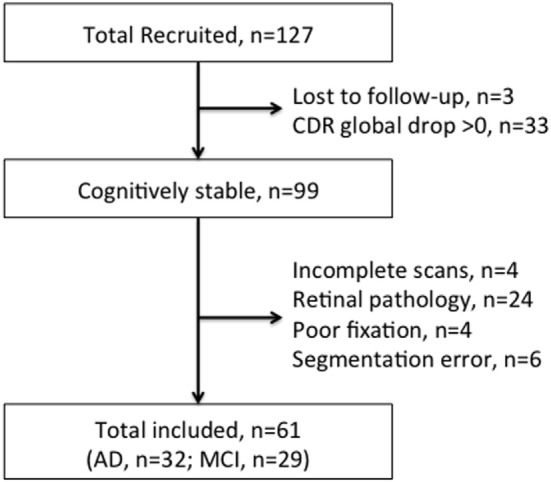
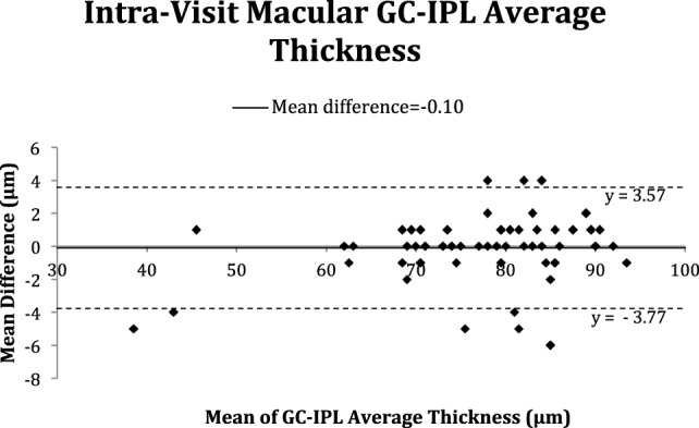
This is a retrospective cohort study. Patients with Alzheimer's disease (AD) or mild cognitive impairment (MCI) with no change in global clinical dementia rating (CDR) score at 1-year follow-up were eligible to be included. Ganglion cell-inner plexiform layer (GC-IPL) and retinal nerve fiber layer (RNFL) parameters were measured with SD-OCT at baseline, 6-month, and 1-year follow-up visits. At baseline, SD-OCT scans were repeated to access intra-visit repeatability of the SD-OCT measurement. SD-OCT measurement over three visits was used to access inter-visit reproducibility. We calculated intraclass correlation coefficients (ICC) and coefficients of variation (CoVs).

RESULTS

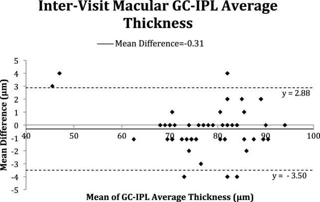
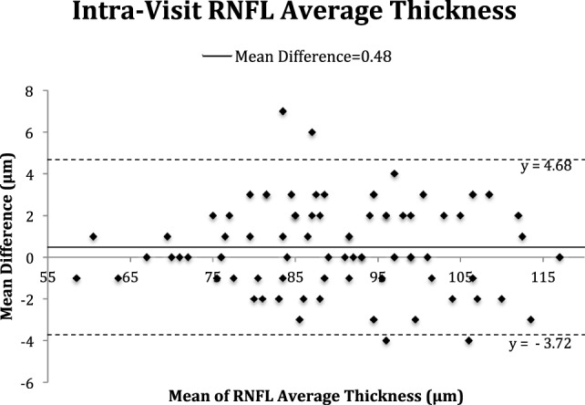
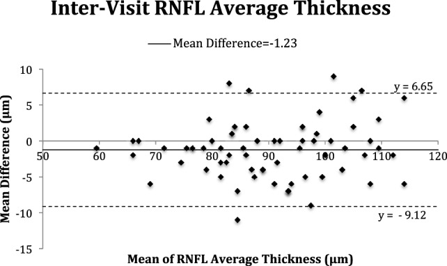
We included 32 patients with stable AD and 29 patients with stable MCI in the final analysis. For GC-IPL measures, the average intra-visit ICC was 0.969 (range: 0.948-0.985), and CoV was 1.81% (range: 1.14-2.40); while the average inter-visit ICC was 0.968 (0.941-0.985), and CoV was 1.91% (range: 1.24-2.32). The average ICC and CoV of intra-visit RNFL measured were 0.965 (range: 0.937-0.986) and 2.32% (range: 1.34-2.90%), respectively. The average ICC and CoV of inter-visit RNFL measures were 0.927 (range: 0.845-0.961) and 3.83% (range: 2.71-5.25%), respectively.

CONCLUSION

Both GC-IPL and RNFL measurements had good intra-visit repeatability and inter-visit reproducibility over 1 year in elderly patients with no decline in cognitive function, suggesting that SD-OCT is a reliable tool to assess neurodegenerative process over time.

摘要

背景

随着人们越来越关注利用光谱域光学相干断层扫描(SD - OCT)测量视网膜神经元结构是否有助于评估认知功能下降和痴呆症发展过程中的神经退行性变,评估SD - OCT测量在这些患者中是否具有可重复性和再现性非常重要。

方法

这是一项回顾性队列研究。纳入在1年随访中全球临床痴呆评定量表(CDR)评分无变化的阿尔茨海默病(AD)或轻度认知障碍(MCI)患者。在基线、6个月和1年随访时用SD - OCT测量神经节细胞 - 内丛状层(GC - IPL)和视网膜神经纤维层(RNFL)参数。在基线时,重复进行SD - OCT扫描以评估SD - OCT测量的访内重复性。三次访视的SD - OCT测量用于评估访间再现性。我们计算了组内相关系数(ICC)和变异系数(CoV)。

结果

最终分析纳入了32例病情稳定的AD患者和29例病情稳定的MCI患者。对于GC - IPL测量,平均访内ICC为0.969(范围:0.948 - 0.985),CoV为1.81%(范围:1.14 - 2.40);而平均访间ICC为0.968(0.941 - 0.985),CoV为1.91%(范围:1.24 - 2.32)。访内RNFL测量的平均ICC和CoV分别为0.965(范围:0.937 - 0.986)和2.32%(范围:1.34 - 2.90%)。访间RNFL测量的平均ICC和CoV分别为0.927(范围:0.845 - 0.961)和3.83%(范围:2.71 - 5.25%)。

结论

在认知功能无下降的老年患者中,GC - IPL和RNFL测量在1年中均具有良好的访内重复性和访间再现性,这表明SD - OCT是评估随时间变化的神经退行性变过程的可靠工具。

https://cdn.ncbi.nlm.nih.gov/pmc/blobs/cddb/5559462/3bebe5fdd57f/fneur-08-00359-g005.jpg
https://cdn.ncbi.nlm.nih.gov/pmc/blobs/cddb/5559462/74a30e0b1a7d/fneur-08-00359-g001.jpg
https://cdn.ncbi.nlm.nih.gov/pmc/blobs/cddb/5559462/7e200c736f20/fneur-08-00359-g002.jpg
https://cdn.ncbi.nlm.nih.gov/pmc/blobs/cddb/5559462/28488ff36102/fneur-08-00359-g003.jpg
https://cdn.ncbi.nlm.nih.gov/pmc/blobs/cddb/5559462/c817849d9900/fneur-08-00359-g004.jpg
https://cdn.ncbi.nlm.nih.gov/pmc/blobs/cddb/5559462/3bebe5fdd57f/fneur-08-00359-g005.jpg

相似文献

1
Repeatability and Reproducibility of Retinal Neuronal and Axonal Measures on Spectral-Domain Optical Coherence Tomography in Patients with Cognitive Impairment.认知障碍患者视网膜神经元和轴突测量在光谱域光学相干断层扫描中的可重复性和再现性
Front Neurol. 2017 Aug 15;8:359. doi: 10.3389/fneur.2017.00359. eCollection 2017.
2
Repeatability of peripapillary retinal nerve fiber layer and inner retinal thickness among two spectral domain optical coherence tomography devices.两种光谱域光学相干断层扫描设备测量视乳头周围视网膜神经纤维层及视网膜内层厚度的可重复性
Invest Ophthalmol Vis Sci. 2014 Sep 16;55(10):6536-46. doi: 10.1167/iovs.14-15072.
3
Retinal ganglion cell analysis using high-definition optical coherence tomography in patients with mild cognitive impairment and Alzheimer's disease.使用高清光学相干断层扫描技术对轻度认知障碍和阿尔茨海默病患者进行视网膜神经节细胞分析。
J Alzheimers Dis. 2015;45(1):45-56. doi: 10.3233/JAD-141659.
4
Repeatability and Reproducibility of Spectral-Domain Optical Coherence Tomography Measurements of Retinal Thickness in Rats.大鼠视网膜厚度的光谱域光学相干断层扫描测量的可重复性和再现性
Curr Eye Res. 2016 Oct;41(10):1346-1352. doi: 10.3109/02713683.2015.1114651. Epub 2016 Feb 10.
5
Comparison of retinal nerve fiber layer measurement between 2 spectral domain OCT instruments.两种谱域光学相干断层扫描仪测量视网膜神经纤维层的比较。
J Glaucoma. 2012 Apr-May;21(4):266-73. doi: 10.1097/IJG.0b013e3182071cdd.
6
Optical Coherence Tomography Reveals Retinal Neuroaxonal Thinning in Frontotemporal Dementia as in Alzheimer's Disease.光学相干断层扫描显示,额颞叶痴呆患者的视网膜神经轴突变薄,与阿尔茨海默病患者情况相同。
J Alzheimers Dis. 2017;56(3):1101-1107. doi: 10.3233/JAD-160886.
7
Repeatability of ganglion cell-inner plexiform layer thickness measurements using spectral-domain OCT in branch retinal vein occlusion.使用光谱域光学相干断层扫描测量视网膜分支静脉阻塞时神经节细胞-内网状层厚度的可重复性
Graefes Arch Clin Exp Ophthalmol. 2017 Sep;255(9):1727-1735. doi: 10.1007/s00417-017-3710-1. Epub 2017 Jun 11.
8
Reliability of Vessel Density Measurements in the Peripapillary Retina and Correlation with Retinal Nerve Fiber Layer Thickness in Healthy Subjects Using Optical Coherence Tomography Angiography.使用光学相干断层扫描血管造影术测量健康受试者视乳头周围视网膜血管密度的可靠性及其与视网膜神经纤维层厚度的相关性
Ophthalmologica. 2018;240(4):183-190. doi: 10.1159/000485957. Epub 2018 Apr 25.
9
Reproducibility of macular, retinal nerve fiber layer, and ONH measurements by OCT in Rhesus monkeys: The Beijing Intracranial and Intraocular Pressure (iCOP) Study.OCT 测量恒河猴眼黄斑、视网膜神经纤维层和视盘的重复性:北京颅内和眼内压(iCOP)研究。
Invest Ophthalmol Vis Sci. 2012 Jul 3;53(8):4505-9. doi: 10.1167/iovs.12-9439.
10
Decreased retinal thickness in patients with Alzheimer's disease is correlated with disease severity.阿尔茨海默病患者的视网膜厚度降低与疾病严重程度相关。
PLoS One. 2019 Nov 5;14(11):e0224180. doi: 10.1371/journal.pone.0224180. eCollection 2019.

引用本文的文献

1
Peripapillary Retinal Nerve Fiber Layer (pRNFL) Thickness - A Novel Biomarker of Neurodegeneration in Late-Infantile CLN2 Disease.视乳头周围视网膜神经纤维层(pRNFL)厚度——晚发性婴儿型CLN2病神经退行性变的一种新型生物标志物。
Eye Brain. 2024 Nov 13;16:101-113. doi: 10.2147/EB.S473408. eCollection 2024.
2
Standardization and clinical applications of retinal imaging biomarkers for cardiovascular disease: a Roadmap from an NHLBI workshop.心血管疾病视网膜成像生物标志物的标准化及临床应用:美国国立心肺血液研究所研讨会路线图
Nat Rev Cardiol. 2025 Jan;22(1):47-63. doi: 10.1038/s41569-024-01060-8. Epub 2024 Jul 22.
3
Altered visual functions, macular ganglion cell and papillary retinal nerve fiber layer thickness in early-treated adult PKU patients.

本文引用的文献

1
Imaging retina to study dementia and stroke.利用影像学检查视网膜以研究痴呆和中风。
Prog Retin Eye Res. 2017 Mar;57:89-107. doi: 10.1016/j.preteyeres.2017.01.001. Epub 2017 Jan 3.
2
A systematic review and meta-analysis of retinal nerve fiber layer change in dementia, using optical coherence tomography.一项使用光学相干断层扫描技术对痴呆患者视网膜神经纤维层变化进行的系统评价和荟萃分析。
Alzheimers Dement (Amst). 2015 Apr 23;1(2):136-43. doi: 10.1016/j.dadm.2015.03.001. eCollection 2015 Jun.
3
The Utilization of Retinal Nerve Fiber Layer Thickness to Predict Cognitive Deterioration.
早期治疗的成年苯丙酮尿症患者的视觉功能、黄斑神经节细胞及视乳头视网膜神经纤维层厚度改变
Mol Genet Metab Rep. 2020 Sep 22;25:100649. doi: 10.1016/j.ymgmr.2020.100649. eCollection 2020 Dec.
4
Retinal axonal degeneration in Niemann-Pick type C disease.尼曼-匹克病 C 型的视网膜轴突变性。
J Neurol. 2020 Jul;267(7):2070-2082. doi: 10.1007/s00415-020-09796-2. Epub 2020 Mar 28.
5
Reflections on the Utility of the Retina as a Biomarker for Alzheimer's Disease: A Literature Review.关于视网膜作为阿尔茨海默病生物标志物的效用的思考:文献综述
Neurol Ther. 2019 Dec;8(Suppl 2):57-72. doi: 10.1007/s40120-019-00173-4. Epub 2019 Dec 12.
6
Validation of Optical Coherence Tomography Retinal Segmentation in Neurodegenerative Disease.光学相干断层扫描视网膜分割在神经退行性疾病中的验证
Transl Vis Sci Technol. 2019 Sep 11;8(5):6. doi: 10.1167/tvst.8.5.6. eCollection 2019 Sep.
7
Identification of Retinal Biomarkers in Alzheimer's Disease Using Optical Coherence Tomography: Recent Insights, Challenges, and Opportunities.利用光学相干断层扫描技术识别阿尔茨海默病中的视网膜生物标志物:最新见解、挑战与机遇
J Clin Med. 2019 Jul 9;8(7):996. doi: 10.3390/jcm8070996.
8
Retinal Nerve Fiber Layer Thinning Is Associated With Brain Atrophy: A Longitudinal Study in Nondemented Older Adults.视网膜神经纤维层变薄与脑萎缩相关:一项针对非痴呆老年人的纵向研究
Front Aging Neurosci. 2019 Apr 11;11:69. doi: 10.3389/fnagi.2019.00069. eCollection 2019.
9
Persistent and Progressive Outer Retina Thinning in Frontotemporal Degeneration.额颞叶痴呆中持续性和进行性的外层视网膜变薄
Front Neurosci. 2019 Apr 4;13:298. doi: 10.3389/fnins.2019.00298. eCollection 2019.
利用视网膜神经纤维层厚度预测认知功能衰退。
J Alzheimers Dis. 2016;49(2):399-405. doi: 10.3233/JAD-150438.
4
Role of Retinal Nerve Fiber Layer Thickness and Optic Disk Measurement by OCT on Early Diagnosis of Glaucoma.光学相干断层扫描测量视网膜神经纤维层厚度和视盘对青光眼早期诊断的作用
Eye Sci. 2015 Mar;30(1):7-12.
5
How strong is the relationship between glaucoma, the retinal nerve fibre layer, and neurodegenerative diseases such as Alzheimer's disease and multiple sclerosis?青光眼、视网膜神经纤维层与阿尔茨海默病和多发性硬化症等神经退行性疾病之间的关系有多紧密?
Eye (Lond). 2015 Oct;29(10):1270-84. doi: 10.1038/eye.2015.158. Epub 2015 Sep 4.
6
Optical Coherence Tomography in Alzheimer's Disease: A Meta-Analysis.光学相干断层扫描技术在阿尔茨海默病中的应用:一项荟萃分析
PLoS One. 2015 Aug 7;10(8):e0134750. doi: 10.1371/journal.pone.0134750. eCollection 2015.
7
Ganglion Cell-Inner Plexiform Layer Thickness in Retinal Diseases: Repeatability Study of Spectral-Domain Optical Coherence Tomography.视网膜疾病中神经节细胞-内网状层厚度:频域光学相干断层扫描的重复性研究
Am J Ophthalmol. 2015 Aug;160(2):283-289.e1. doi: 10.1016/j.ajo.2015.05.015. Epub 2015 May 21.
8
Long-Term Reproducibility of Macular Ganglion Cell Analysis in Clinically Stable Glaucoma Patients.临床稳定青光眼患者黄斑神经节细胞分析的长期可重复性
Invest Ophthalmol Vis Sci. 2015 Jul;56(8):4857-64. doi: 10.1167/iovs.14-16350.
9
Test-retest variability of retinal nerve fiber layer thickness and macular ganglion cell-inner plexiform layer thickness measurements using spectral-domain optical coherence tomography.使用频域光学相干断层扫描技术测量视网膜神经纤维层厚度和黄斑神经节细胞-内丛状层厚度的重测变异性。
J Glaucoma. 2015 Jun-Jul;24(5):e109-15. doi: 10.1097/IJG.0000000000000203.
10
Retinal neurodegeneration on optical coherence tomography and cerebral atrophy.光学相干断层扫描显示的视网膜神经变性和脑萎缩。
Neurosci Lett. 2015 Jan 1;584:12-6. doi: 10.1016/j.neulet.2014.10.010. Epub 2014 Oct 19.