Olszewska Agnieszka, Farke Daniela, Schmidt Martin Jürgen
Department of Veterinary Clinical Sciences, Small Animal Clinic, Neurosurgery, Neuroradiology and Clinical Neurology, Justus-Liebig-University, Frankfurter Strasse 108, 35392, Giessen, Germany.
Ir Vet J. 2020 Mar 25;73:5. doi: 10.1186/s13620-020-00159-x. eCollection 2020.
Overdrainage and collapse of the hemispheres is a potential severe complication after surgical treatment of internal hydrocephalus using ventriculoperitoneal shunts. Here we describe a case of a spontaneous hemispheric ventricular collapse in an untreated dog with congenital hydrocephalus internus.
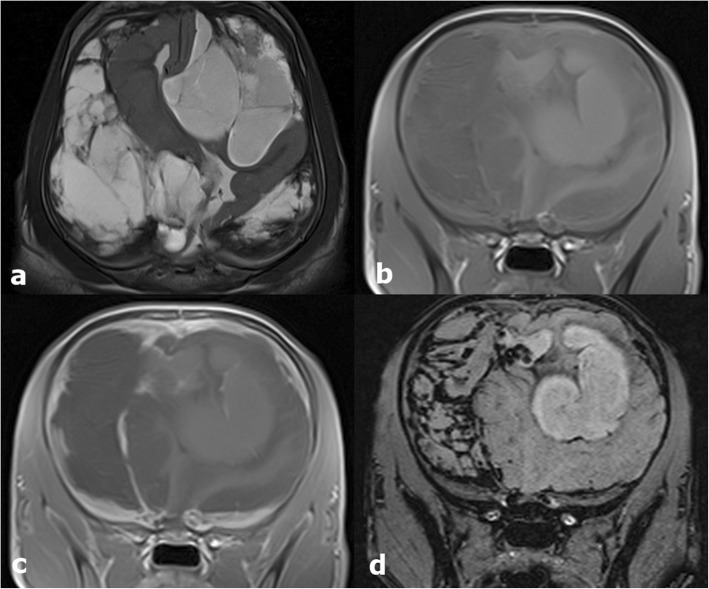
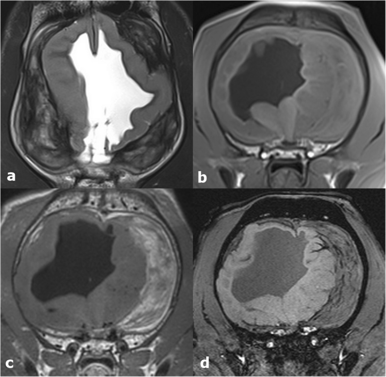
A twelve-week-old, male, intact Golden Retriever was presented with a history of peracute obtundation, impaired vision, and progressive gait abnormalities of all limbs for three days. Neurological examination revealed a dome shaped skull, a broad-based stance and a moderate cerebellar ataxia. The postural responses were markedly delayed in all limbs. Moderate ventro-lateral strabismus, vertical nystagmus and absent menace response were observed bilaterally. Clinical signs indicated multifocal localisation (forebrain, cerebellum). Magnetic resonance imaging (MRI) showed dilation of all cerebral ventricles, irregular thinning of the periventricular white and grey matter, consistent with internal hydrocephalus. In addition, the hemispheres were collapsed at the right temporal and left frontal lobe with haemorrhage filling the adjacent subarachnoid space. The dog underwent left frontal and right temporal craniotomy for removal of the haemorrhage. The dog improved on all neurological signs and was discharged after seven days. A repeat MRI three months postsurgical intervention showed reexpansion of the cerebral hemispheres. Subarachnoid haemorrhages were markedly reduced.
Collapse of the hemispheres can occur spontaneously in dogs with hydrocephalus internus. Removal of the haemorrhage can improve clinical signs.
使用脑室腹腔分流术治疗内部脑积水后,半球过度引流和塌陷是一种潜在的严重并发症。在此,我们描述一例未经治疗的先天性内部脑积水犬自发性半球脑室塌陷的病例。
一只12周龄、雄性、未绝育的金毛寻回犬,有急性昏迷、视力受损和四肢进行性步态异常3天的病史。神经学检查发现颅骨呈圆顶状、宽基步态和中度小脑共济失调。所有肢体的姿势反应明显延迟。双侧观察到中度腹外侧斜视、垂直眼球震颤和威胁反应消失。临床症状表明存在多灶性定位(前脑、小脑)。磁共振成像(MRI)显示所有脑室扩张,脑室周围白质和灰质不规则变薄,符合内部脑积水。此外,右颞叶和左额叶的半球塌陷,出血填充相邻的蛛网膜下腔。该犬接受了左额叶和右颞叶开颅手术以清除出血。犬的所有神经症状均有改善,7天后出院。术后3个月的重复MRI显示大脑半球重新扩张。蛛网膜下腔出血明显减少。
内部脑积水犬可发生自发性半球塌陷。清除出血可改善临床症状。