Hashemzadeh Hadi, Allahverdi Abdollah, Sedghi Mosslim, Vaezi Zahra, Tohidi Moghadam Tahereh, Rothbauer Mario, Fischer Michael Bernhard, Ertl Peter, Naderi-Manesh Hossein
Department of Nanobiotechnology, Faculty of Biological Science, Tarbiat Modares University, Tehran 14115-154, Iran.
Department of Biophysics, Faculty of Biological Science, Tarbiat Modares University, Tehran 14115-154, Iran.
Nanomaterials (Basel). 2020 Apr 2;10(4):668. doi: 10.3390/nano10040668.
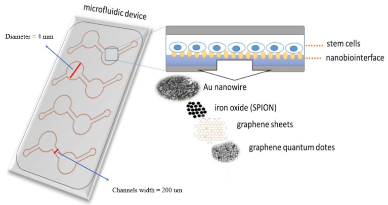
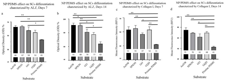
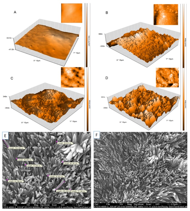
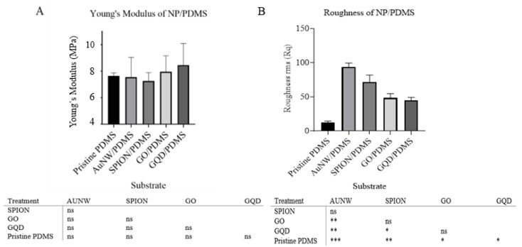
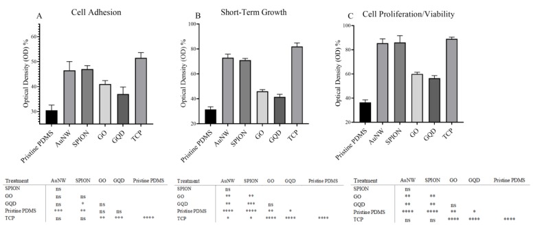
Microfluidics cell-based assays require strong cell-substrate adhesion for cell viability, proliferation, and differentiation. The intrinsic properties of PDMS, a commonly used polymer in microfluidics systems, regarding cell-substrate interactions have limited its application for microfluidics cell-based assays. Various attempts by previous researchers, such as chemical modification, plasma-treatment, and protein-coating of PDMS revealed some improvements. These strategies are often reversible, time-consuming, short-lived with either cell aggregates formation, not cost-effective as well as not user- and eco-friendly too. To address these challenges, cell-surface interaction has been tuned by the modification of PDMS doped with different biocompatible nanomaterials. Gold nanowires (AuNWs), superparamagnetic iron oxide nanoparticles (SPIONs), graphene oxide sheets (GO), and graphene quantum dot (GQD) have already been coupled to PDMS as an alternative biomaterial enabling easy and straightforward integration during microfluidic fabrication. The synthesized nanoparticles were characterized by corresponding methods. Physical cues of the nanostructured substrates such as Young's modulus, surface roughness, and nanotopology have been carried out using atomic force microscopy (AFM). Initial biocompatibility assessment of the nanocomposites using human amniotic mesenchymal stem cells (hAMSCs) showed comparable cell viabilities among all nanostructured PDMS composites. Finally, osteogenic stem cell differentiation demonstrated an improved differentiation rate inside microfluidic devices. The results revealed that the presence of nanomaterials affected a 5- to 10-fold increase in surface roughness. In addition, the results showed enhancement of cell proliferation from 30% (pristine PDMS) to 85% (nano-modified scaffolds containing AuNWs and SPIONs), calcification from 60% (pristine PDMS) to 95% (PDMS/AuNWs), and cell surface marker expression from 40% in PDMS to 77% in SPION- and AuNWs-PDMS scaffolds at 14 day. Our results suggest that nanostructured composites have a very high potential for stem cell studies and future therapies.
基于微流控芯片的细胞分析需要强大的细胞与基质粘附力以维持细胞活力、增殖和分化。聚二甲基硅氧烷(PDMS)是微流控系统中常用的聚合物,其在细胞与基质相互作用方面的固有特性限制了它在基于微流控芯片的细胞分析中的应用。先前研究人员进行了各种尝试,如对PDMS进行化学修饰、等离子体处理和蛋白质包被,取得了一些改进。但这些策略往往具有可逆性、耗时、寿命短,还会形成细胞聚集体,既不具有成本效益,也不便于用户操作且不环保。为应对这些挑战,通过掺杂不同生物相容性纳米材料对PDMS进行改性来调节细胞表面相互作用。金纳米线(AuNWs)、超顺磁性氧化铁纳米颗粒(SPIONs)、氧化石墨烯片(GO)和石墨烯量子点(GQD)已被与PDMS偶联,作为一种替代生物材料,便于在微流控制造过程中轻松直接地集成。通过相应方法对合成的纳米颗粒进行了表征。使用原子力显微镜(AFM)对纳米结构基质的物理线索,如杨氏模量、表面粗糙度和纳米拓扑结构进行了研究。使用人羊膜间充质干细胞(hAMSCs)对纳米复合材料进行的初步生物相容性评估表明,所有纳米结构的PDMS复合材料的细胞活力相当。最后,成骨干细胞分化显示微流控装置内的分化率有所提高。结果表明,纳米材料的存在使表面粗糙度增加了5到10倍。此外,结果显示细胞增殖从30%(原始PDMS)提高到85%(含有AuNWs和SPIONs的纳米改性支架),钙化从60%(原始PDMS)提高到95%(PDMS/AuNWs),细胞表面标志物表达在第14天从PDMS中的40%提高到SPIONs和AuNWs - PDMS支架中的77%。我们的结果表明,纳米结构复合材料在干细胞研究和未来治疗方面具有很高的潜力。