• 文献检索
  • 文档翻译
  • 深度研究
  • 学术资讯
  • Suppr Zotero 插件Zotero 插件
  • 邀请有礼
  • 套餐&价格
  • 历史记录
应用&插件
Suppr Zotero 插件Zotero 插件浏览器插件Mac 客户端Windows 客户端微信小程序
定价
高级版会员购买积分包购买API积分包
服务
文献检索文档翻译深度研究API 文档MCP 服务
关于我们
关于 Suppr公司介绍联系我们用户协议隐私条款
关注我们

Suppr 超能文献

核心技术专利:CN118964589B侵权必究
粤ICP备2023148730 号-1Suppr @ 2026

文献检索

告别复杂PubMed语法,用中文像聊天一样搜索,搜遍4000万医学文献。AI智能推荐,让科研检索更轻松。

立即免费搜索

文件翻译

保留排版,准确专业,支持PDF/Word/PPT等文件格式,支持 12+语言互译。

免费翻译文档

深度研究

AI帮你快速写综述,25分钟生成高质量综述,智能提取关键信息,辅助科研写作。

立即免费体验

渗透压和氧张力对软骨组织工程的重要性。

Importance of Osmolarity and Oxygen Tension for Cartilage Tissue Engineering.

作者信息

Sieber Stefan, Michaelis Martin, Gühring Hans, Lindemann Sven, Gigout Anne

机构信息

Osteoarthritis Research, Merck KGaA, Darmstadt, Germany.

出版信息

Biores Open Access. 2020 Mar 31;9(1):106-115. doi: 10.1089/biores.2020.0009. eCollection 2020.

DOI:10.1089/biores.2020.0009
PMID:32257626
原文链接:https://pmc.ncbi.nlm.nih.gov/articles/PMC7133430/
Abstract

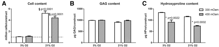
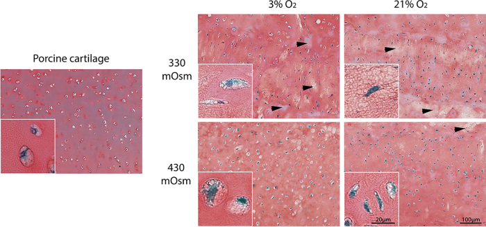
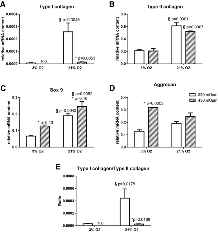
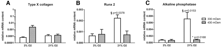
For cartilage repair or evaluation of new therapeutic approaches , the generation of functional cartilage tissue is of crucial importance and can only be achieved if the phenotype of the chondrocytes is preserved. Three-dimensional (3D) cell culture is broadly used for this purpose. However, adapting culture parameters like the oxygen tension or the osmolarity to their physiological values is often omitted. Indeed, articular cartilage is an avascular tissue subjected to reduced oxygen tension and presenting and increased osmolarity compared with most other tissues. In this study, we aimed at evaluating the effect of a physiological oxygen tension (3% instead of 21%) and physiological osmolarity (430 vs. 330 mOsm in nonadjusted DMEM) and the combination of both on the cell proliferation, matrix production, and the phenotype of porcine chondrocytes in a scaffold-free 3D culture system. We observed that a physiological osmolarity had no effect on cell proliferation and matrix production but positively influences the chondrocyte phenotype. A physiological oxygen level prevented cell proliferation but resulted in an increased matrix content/million cells and had a positive influence on the chondrocyte phenotype as well. The strongest benefit was reached with the combination of both physiological osmolarity and oxygen levels; with these conditions, type I collagen expression became undetectable. In addition, at 3% O the chondrocytes-matrix constructs were found to more closely resemble native cartilage regarding the matrix-to-cell ratio. In conclusion, this study clearly demonstrates the benefit of using physiological oxygen tension and osmolarity in cartilage tissue engineering with the combination of both showing the strongest benefit on the chondrocyte phenotype.

摘要

对于软骨修复或新治疗方法的评估而言,功能性软骨组织的生成至关重要,并且只有在软骨细胞表型得以保留的情况下才能实现。三维(3D)细胞培养广泛用于此目的。然而,常常忽略将培养参数如氧张力或渗透压调整至其生理值。实际上,与大多数其他组织相比,关节软骨是一种无血管组织,其氧张力降低且渗透压升高。在本研究中,我们旨在评估生理氧张力(3%而非21%)、生理渗透压(未调整的DMEM中为430与330 mOsm)以及二者组合对无支架3D培养系统中猪软骨细胞的细胞增殖、基质产生和表型的影响。我们观察到生理渗透压对细胞增殖和基质产生没有影响,但对软骨细胞表型有积极影响。生理氧水平可阻止细胞增殖,但导致每百万细胞的基质含量增加,并且对软骨细胞表型也有积极影响。生理渗透压和氧水平二者组合带来的益处最为显著;在这些条件下,I型胶原蛋白表达变得不可检测。此外,在3% O₂时,软骨细胞 - 基质构建体在基质与细胞比例方面被发现更类似于天然软骨。总之,本研究清楚地证明了在软骨组织工程中使用生理氧张力和渗透压的益处,二者组合对软骨细胞表型显示出最强的益处。

https://cdn.ncbi.nlm.nih.gov/pmc/blobs/b40d/7133430/4318f91d27ab/biores.2020.0009_figure5.jpg
https://cdn.ncbi.nlm.nih.gov/pmc/blobs/b40d/7133430/58a484783839/biores.2020.0009_figure1.jpg
https://cdn.ncbi.nlm.nih.gov/pmc/blobs/b40d/7133430/55251e596e91/biores.2020.0009_figure2.jpg
https://cdn.ncbi.nlm.nih.gov/pmc/blobs/b40d/7133430/1c7ff3555fb3/biores.2020.0009_figure3.jpg
https://cdn.ncbi.nlm.nih.gov/pmc/blobs/b40d/7133430/78f6ccfa6628/biores.2020.0009_figure4.jpg
https://cdn.ncbi.nlm.nih.gov/pmc/blobs/b40d/7133430/4318f91d27ab/biores.2020.0009_figure5.jpg

相似文献

1
Importance of Osmolarity and Oxygen Tension for Cartilage Tissue Engineering.渗透压和氧张力对软骨组织工程的重要性。
Biores Open Access. 2020 Mar 31;9(1):106-115. doi: 10.1089/biores.2020.0009. eCollection 2020.
2
Synoviocyte Derived-Extracellular Matrix Enhances Human Articular Chondrocyte Proliferation and Maintains Re-Differentiation Capacity at Both Low and Atmospheric Oxygen Tensions.滑膜细胞衍生的细胞外基质在低氧和常压氧张力下均能增强人关节软骨细胞的增殖并维持再分化能力。
PLoS One. 2015 Jun 15;10(6):e0129961. doi: 10.1371/journal.pone.0129961. eCollection 2015.
3
Osmolarity modulates the de-differentiation of horse articular chondrocytes during cell expansion in vitro: implications for tissue engineering in cartilage repair.渗透压调节体外细胞扩增过程中马关节软骨细胞去分化:对软骨修复组织工程的影响。
Vet Res Commun. 2023 Dec;47(4):2285-2292. doi: 10.1007/s11259-023-10140-y. Epub 2023 May 19.
4
Physiological osmolarities do not enhance long-term tissue synthesis in chondrocyte-laden degradable poly(ethylene glycol) hydrogels.生理渗透压不会增强负载软骨细胞的可降解聚乙二醇水凝胶中的长期组织合成。
J Biomed Mater Res A. 2015 Jun;103(6):2186-92. doi: 10.1002/jbm.a.35329. Epub 2014 Sep 24.
5
Increased TGF-β and BMP Levels and Improved Chondrocyte-Specific Marker Expression In Vitro under Cartilage-Specific Physiological Osmolarity.在软骨特异性生理渗透压下,TGF-β和 BMP 水平升高,软骨细胞特异性标志物表达改善。
Int J Mol Sci. 2019 Feb 13;20(4):795. doi: 10.3390/ijms20040795.
6
Disparate response of articular- and auricular-derived chondrocytes to oxygen tension.关节和耳源软骨细胞对氧张力的不同反应。
Connect Tissue Res. 2016 Jul;57(4):319-33. doi: 10.1080/03008207.2016.1182996. Epub 2016 Apr 29.
7
Effect of low oxygen tension on tissue-engineered cartilage construct development in the concentric cylinder bioreactor.低氧张力对同心圆柱生物反应器中组织工程软骨构建体发育的影响。
Tissue Eng. 2004 May-Jun;10(5-6):825-32. doi: 10.1089/1076327041348545.
8
Osmolarity influences chondrocyte death in wounded articular cartilage.渗透压影响创伤性关节软骨中的软骨细胞死亡。
J Bone Joint Surg Am. 2008 Jul;90(7):1531-42. doi: 10.2106/JBJS.G.00857.
9
Hypertonic conditions enhance cartilage formation in scaffold-free primary chondrocyte cultures.高渗条件可增强无支架原代软骨细胞培养中的软骨形成。
Cell Tissue Res. 2014 Nov;358(2):541-50. doi: 10.1007/s00441-014-1970-1. Epub 2014 Aug 9.
10
Redifferentiation of in vitro expanded adult articular chondrocytes by combining the hanging-drop cultivation method with hypoxic environment.通过将悬滴培养法与低氧环境相结合实现体外扩增的成年关节软骨细胞的再分化。
Cell Transplant. 2008;17(8):987-96. doi: 10.3727/096368908786576499.

引用本文的文献

1
Co-culture engineering: a promising strategy for production of engineered extracellular vesicle for osteoarthritis treatment.共培养工程:一种有前途的策略,用于生产用于骨关节炎治疗的工程细胞外囊泡。
Cell Commun Signal. 2024 Jan 10;22(1):29. doi: 10.1186/s12964-023-01467-9.
2
Membrane stretch as the mechanism of activation of PIEZO1 ion channels in chondrocytes.细胞膜拉伸作为软骨细胞中 PIEZO1 离子通道激活的机制。
Proc Natl Acad Sci U S A. 2023 Jul 25;120(30):e2221958120. doi: 10.1073/pnas.2221958120. Epub 2023 Jul 17.
3
Investigating the interfaces of the epiphyseal plate: An integrated approach of histochemistry, microtomography and SEM.

本文引用的文献

1
Tissue-engineered cartilage: the crossroads of biomaterials, cells and stimulating factors.组织工程软骨:生物材料、细胞与刺激因子的交叉领域
Macromol Biosci. 2015 Feb;15(2):153-82. doi: 10.1002/mabi.201400335. Epub 2014 Oct 3.
2
Repair and tissue engineering techniques for articular cartilage.关节软骨的修复与组织工程技术
Nat Rev Rheumatol. 2015 Jan;11(1):21-34. doi: 10.1038/nrrheum.2014.157. Epub 2014 Sep 23.
3
Hypertonic conditions enhance cartilage formation in scaffold-free primary chondrocyte cultures.高渗条件可增强无支架原代软骨细胞培养中的软骨形成。
研究骨骺板界面:组织化学、显微断层扫描和扫描电子显微镜的综合方法。
J Anat. 2023 Nov;243(5):870-877. doi: 10.1111/joa.13924. Epub 2023 Jun 30.
4
O variant chip to simulate site-specific skeletogenesis from hypoxic bone marrow.模拟缺氧骨髓中特定部位骨生成的变异芯片。
Sci Adv. 2023 Mar 22;9(12):eadd4210. doi: 10.1126/sciadv.add4210.
5
Engineering Inflammation-Resistant Cartilage: Bridging Gene Therapy and Tissue Engineering.工程化抗炎症软骨:基因治疗与组织工程的桥梁。
Adv Healthc Mater. 2023 Jul;12(17):e2202271. doi: 10.1002/adhm.202202271. Epub 2023 Mar 15.
6
A Progress Report and Roadmap for Microphysiological Systems and Organ-On-A-Chip Technologies to Be More Predictive Models in Human (Knee) Osteoarthritis.微生理系统和器官芯片技术在人类(膝关节)骨关节炎中成为更具预测性模型的进展报告与路线图
Front Bioeng Biotechnol. 2022 Jun 15;10:886360. doi: 10.3389/fbioe.2022.886360. eCollection 2022.
7
Synoviocyte-Derived Extracellular Matrix and bFGF Speed Human Chondrocyte Proliferation While Maintaining Differentiation Potential.滑膜细胞衍生的细胞外基质和碱性成纤维细胞生长因子可加速人软骨细胞增殖,同时维持其分化潜能。
Front Bioeng Biotechnol. 2022 May 24;10:825005. doi: 10.3389/fbioe.2022.825005. eCollection 2022.
8
Physosmotic Induction of Chondrogenic Maturation Is TGF-β Dependent and Enhanced by Calcineurin Inhibitor FK506.物理渗透诱导软骨成熟依赖于 TGF-β 并可被钙调神经磷酸酶抑制剂 FK506 增强。
Int J Mol Sci. 2022 May 4;23(9):5110. doi: 10.3390/ijms23095110.
9
Infrapatellar fat pad adipose-derived stem cells co-cultured with articular chondrocytes from osteoarthritis patients exhibit increased chondrogenic gene expression.髌下脂肪垫脂肪来源干细胞与骨关节炎患者关节软骨细胞共培养后,软骨基因表达增加。
Cell Commun Signal. 2022 Feb 12;20(1):17. doi: 10.1186/s12964-021-00815-x.
10
Scaffold-Based Tissue Engineering Strategies for Osteochondral Repair.用于骨软骨修复的基于支架的组织工程策略。
Front Bioeng Biotechnol. 2022 Jan 11;9:812383. doi: 10.3389/fbioe.2021.812383. eCollection 2021.
Cell Tissue Res. 2014 Nov;358(2):541-50. doi: 10.1007/s00441-014-1970-1. Epub 2014 Aug 9.
4
Hypoxia promotes redifferentiation and suppresses markers of hypertrophy and degeneration in both healthy and osteoarthritic chondrocytes.缺氧可促进健康软骨细胞和骨关节炎软骨细胞的再分化,并抑制肥大和退变标志物。
Arthritis Res Ther. 2013 Aug 21;15(4):R92. doi: 10.1186/ar4272.
5
Multimodal evaluation of tissue-engineered cartilage.组织工程软骨的多模态评估
J Med Biol Eng. 2013 Feb 1;33(1):1-16. doi: 10.5405/jmbe.1254.
6
Osmolarity determines the in vitro chondrogenic differentiation capacity of progenitor cells via nuclear factor of activated T-cells 5.渗透压通过激活 T 细胞核因子 5 决定祖细胞的体外软骨分化能力。
Bone. 2013 Mar;53(1):94-102. doi: 10.1016/j.bone.2012.11.032. Epub 2012 Dec 4.
7
Five percent oxygen tension is not beneficial for neocartilage formation in scaffold-free cell cultures.5%氧气张力不利于无支架细胞培养中成软骨形成。
Cell Tissue Res. 2012 Apr;348(1):109-17. doi: 10.1007/s00441-012-1366-z. Epub 2012 Mar 6.
8
Inhibiting calcineurin activity under physiologic tonicity elevates anabolic but suppresses catabolic chondrocyte markers.在生理张力下抑制钙调神经磷酸酶活性可提高合成代谢,但会抑制分解代谢的软骨细胞标志物。
Arthritis Rheum. 2012 Jun;64(6):1929-39. doi: 10.1002/art.34369. Epub 2012 Jan 9.
9
Chondrocyte hypertrophy and osteoarthritis: role in initiation and progression of cartilage degeneration?软骨细胞肥大和骨关节炎:在软骨退变的起始和进展中的作用?
Osteoarthritis Cartilage. 2012 Mar;20(3):223-32. doi: 10.1016/j.joca.2011.12.003. Epub 2011 Dec 11.
10
Combined 3D and hypoxic culture improves cartilage-specific gene expression in human chondrocytes.三维共培养和低氧培养提高人软骨细胞的软骨特异性基因表达。
Acta Orthop. 2011 Apr;82(2):234-40. doi: 10.3109/17453674.2011.566135. Epub 2011 Mar 24.