Department of Anatomy and Cell Biology, University Hospital RWTH Aachen University, 52074 Aachen, Germany.
Department of Orthopaedic Surgery, Maastricht University Medical Center, 6229 HX Maastricht, The Netherlands.
Int J Mol Sci. 2022 May 4;23(9):5110. doi: 10.3390/ijms23095110.
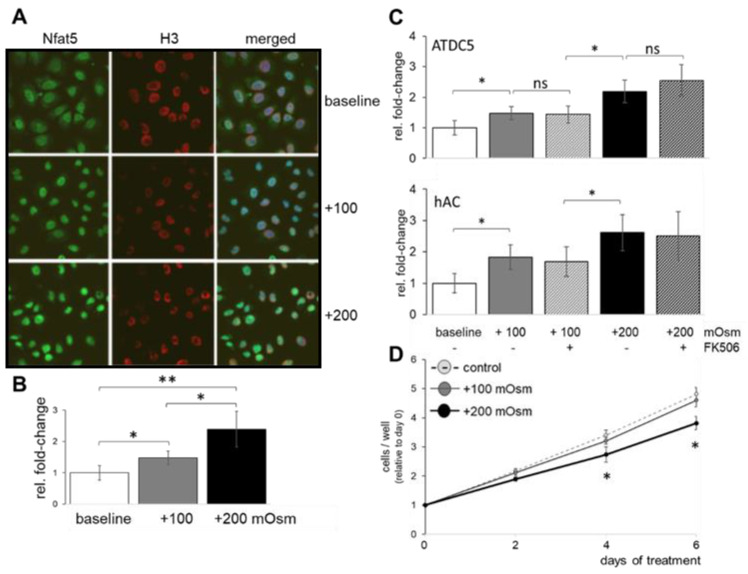
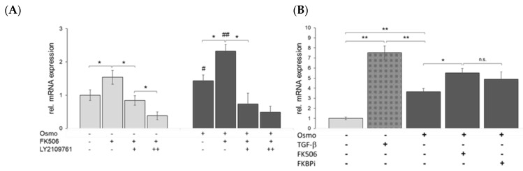
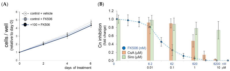
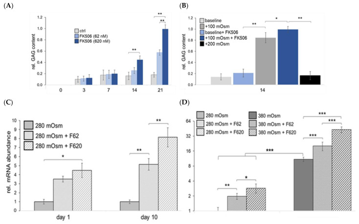
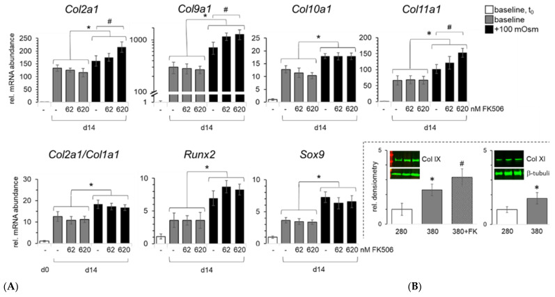
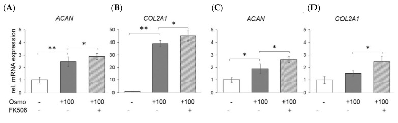
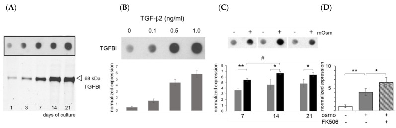
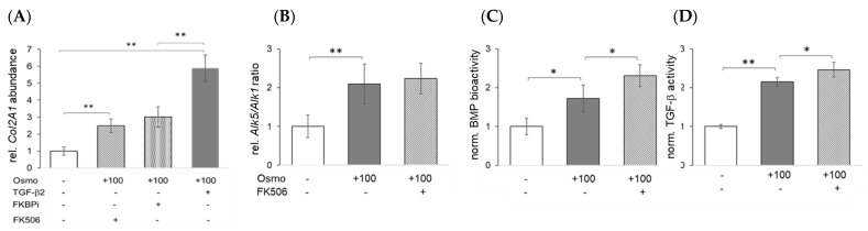
Increasing extracellular osmolarity 100 mOsm/kg above plasma level to the physiological levels for cartilage induces chondrogenic marker expression and the differentiation of chondroprogenitor cells. The calcineurin inhibitor FK506 has been reported to modulate the hypertrophic differentiation of primary chondrocytes under such conditions, but the molecular mechanism has remained unclear. We aimed at clarifying its role. Chondrocyte cell lines and primary cells were cultured under plasma osmolarity and chondrocyte-specific in situ osmolarity (+100 mOsm, physosmolarity) was increased to compare the activation of nuclear factor of activated T-cells 5 (NFAT5). The effects of osmolarity and FK506 on calcineurin activity, cell proliferation, extracellular matrix quality, and BMP- and TGF-β signaling were analyzed using biochemical, gene, and protein expression, as well as reporter and bio-assays. NFAT5 translocation was similar in chondrocyte cell lines and primary cells. High supraphysiological osmolarity compromised cell proliferation, while physosmolarity or FK506 did not, but in combination increased proteoglycan and collagen expression in chondrocytes in vitro and in situ. The expression of the TGF-β-inducible protein TGFBI, as well as chondrogenic (, and terminal differentiation markers (e.g., ) were affected by osmolarity. Particularly, the expression of minor collagens (e.g., , ) was affected. The inhibition of the FK506-binding protein suggests modulation at the TGF-β receptor level, rather than calcineurin-mediated signaling, as a cause. Physiological osmolarity promotes terminal chondrogenic differentiation of progenitor cells through the sensitization of the TGF-β superfamily signaling at the type I receptor. While hyperosmolarity alone facilitates TGF-β superfamily signaling, FK506 further enhances signaling by releasing the FKBP12 break from the type I receptor to improve collagenous marker expression. Our results help explain earlier findings and potentially benefit future cell-based cartilage repair strategies.
将细胞外渗透压提高到比血浆水平高 100mOsm/kg 的生理水平可诱导软骨细胞的软骨形成标志物表达和软骨祖细胞的分化。已经报道钙调神经磷酸酶抑制剂 FK506 可在这种条件下调节原代软骨细胞的肥大分化,但分子机制尚不清楚。我们旨在阐明其作用。在血浆渗透压下培养软骨细胞系和原代细胞,并增加软骨细胞特异性原位渗透压(+100mOsm,生理渗透压)以比较激活 T 细胞活化核因子 5(NFAT5)。使用生化、基因和蛋白质表达以及报告基因和生物测定法分析渗透压和 FK506 对钙调神经磷酸酶活性、细胞增殖、细胞外基质质量以及 BMP 和 TGF-β信号的影响。NFAT5 易位在软骨细胞系和原代细胞中相似。高渗渗透压会损害细胞增殖,而生理渗透压或 FK506 则不会,但会在体外和原位增加软骨细胞中的蛋白聚糖和胶原蛋白表达。TGF-β诱导蛋白 TGFBI 的表达以及软骨形成(、和终末分化标志物(例如、)受渗透压影响。特别是,较小的胶原蛋白(例如、)的表达受到影响。FK506 结合蛋白的抑制表明,这种影响是在 TGF-β受体水平上而非钙调神经磷酸酶介导的信号转导上发生的,因为 FK506 结合蛋白的抑制提示 FK506 结合蛋白在 TGF-β 受体水平上发生了调节,而不是钙调神经磷酸酶介导的信号转导。生理渗透压通过在 I 型受体上敏化 TGF-β 超家族信号来促进祖细胞的终末软骨分化。虽然高渗渗透压本身可促进 TGF-β 超家族信号,但 FK506 通过从 I 型受体释放 FKBP12 阻断物进一步增强信号,从而改善胶原蛋白标志物的表达。我们的结果有助于解释早期的发现,并可能有益于未来基于细胞的软骨修复策略。