West Virginia University Heart and Vascular Institute, 1 Medical Center Drive, Morgantown, WV 26506, USA.
West Virginia University Heart and Vascular Institute, 1 Medical Center Drive, Morgantown, WV 26506, USA.
EBioMedicine. 2020 Apr;54:102726. doi: 10.1016/j.ebiom.2020.102726. Epub 2020 Apr 6.
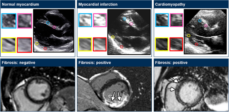
Maturation of ultrasound myocardial tissue characterization may have far-reaching implications as a widely available alternative to cardiac magnetic resonance (CMR) for risk stratification in left ventricular (LV) remodeling.
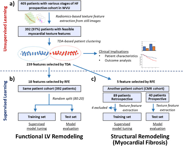
We extracted 328 texture-based features of myocardium from still ultrasound images. After we explored the phenotypes of myocardial textures using unsupervised similarity networks, global LV remodeling parameters were predicted using supervised machine learning models. Separately, we also developed supervised models for predicting the presence of myocardial fibrosis using another cohort who underwent cardiac magnetic resonance (CMR). For the prediction, patients were divided into a training and test set (80:20).
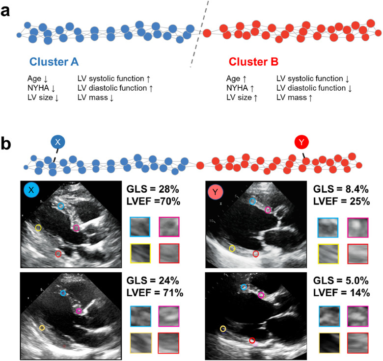
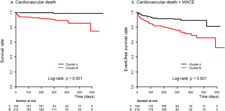
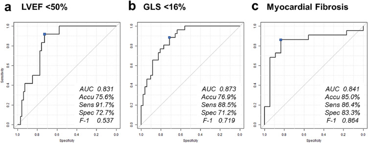
Texture-based tissue feature extraction was feasible in 97% of total 534 patients. Interpatient similarity analysis delineated two patient groups based on the texture features: one group had more advanced LV remodeling parameters compared to the other group. Furthermore, this group was associated with a higher incidence of cardiac deaths (p = 0.001) and major adverse cardiac events (p < 0.001). The supervised models predicted reduced LV ejection fraction (<50%) and global longitudinal strain (<16%) with area under the receiver-operator-characteristics curves (ROC AUC) of 0.83 and 0.87 in the hold-out test set, respectively. Furthermore, the presence of myocardial fibrosis was predicted from only ultrasound myocardial texture with an ROC AUC of 0.84 (sensitivity 86.4% and specificity 83.3%) in the test set.
Ultrasound texture-based myocardial tissue characterization identified phenotypic features of LV remodeling from still ultrasound images. Further clinical validation may address critical barriers in the adoption of ultrasound techniques for myocardial tissue characterization.
None.
超声心肌组织特征成熟可能具有深远的意义,因为它作为一种广泛可用的替代心脏磁共振(CMR)的方法,可用于左心室(LV)重构的风险分层。
我们从静态超声图像中提取了 328 种基于纹理的心肌特征。在使用无监督相似性网络探索心肌纹理表型后,使用监督机器学习模型预测整体 LV 重构参数。另外,我们还使用另一批接受心脏磁共振(CMR)检查的患者开发了用于预测心肌纤维化存在的监督模型。对于预测,患者被分为训练集和测试集(80:20)。
在总共 534 例患者中,有 97%的患者可以进行基于纹理的组织特征提取。患者间相似性分析根据纹理特征将患者分为两组:一组与另一组相比,LV 重构参数更为先进。此外,该组与较高的心脏死亡发生率(p=0.001)和主要不良心脏事件(p<0.001)相关。在保留测试集中,监督模型预测左心室射血分数(<50%)和整体纵向应变(<16%)的受试者工作特征曲线下面积(ROC AUC)分别为 0.83 和 0.87。此外,仅使用超声心肌纹理就可以预测心肌纤维化的存在,在测试集中的 ROC AUC 为 0.84(灵敏度 86.4%,特异性 83.3%)。
基于超声的心肌组织特征纹理分析从静态超声图像中识别出 LV 重构的表型特征。进一步的临床验证可能会解决在采用超声技术进行心肌组织特征分析方面的关键障碍。
无。