Department of Radiology, Trakya University School of Medicine, Edirne, Turkey
Department of Biostatistics and Medical Informatics, Trakya University School of Medicine, Edirne, Turkey
Balkan Med J. 2020 Jun 1;37(4):203-207. doi: 10.4274/balkanmedj.galenos.2020.2019.11.91. Epub 2020 Apr 9.
Primary Sjögren’s syndrome is a chronic inflammatory autoimmune disease. Minor salivary gland biopsy is the gold standard for the diagnosis of primary Sjögren’s syndrome. Superb microvascular imaging, power Doppler ultrasound, and color Doppler of the salivary glands represent non-invasive, non-irradiating modality for evaluating the vascularity of the salivary glands in the diagnosis and follow-up of primary Sjögren’s syndrome.
To evaluate the efficacy of superb microvascular imaging and vascularity index in salivary glands for the sonographic diagnosis of primary Sjögren’s syndrome.
Prospective case-control study.
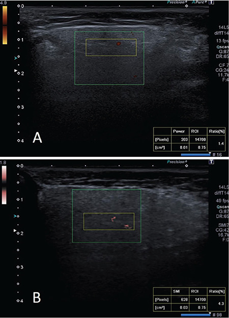
Twenty participants with primary Sjögren’s syndrome and 20 healthy subjects were included in the study. Both parotid glands and submandibular glands were evaluated by superb microvascular imaging, power Doppler ultrasound, and color Doppler. The diagnostic accuracy of superb microvascular imaging was compared using these techniques.
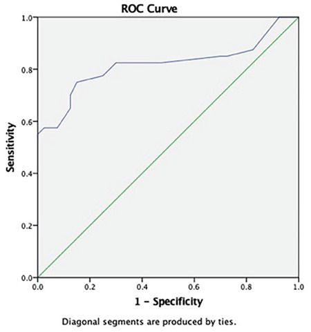
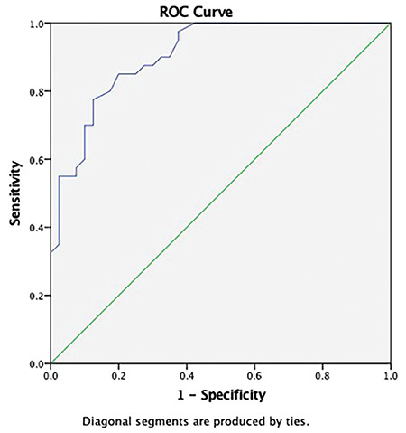
In the patient group, the vascularity index values of superb microvascular imaging in parotid glands and submandibular glands were 3.5±1.66, 5.06±1.94, respectively. While the same values were 1.0±0.98 and 2.44±1.34 in the control group (p≤0.001). In the patient group, the vascularity index values of power Doppler ultrasound in parotid glands and submandibular glands were 1.3±1.20 and 2.59±1.82, respectively. While the same values were 0.3±0.32 and 0.85±0.68 in the control group (p≤0.001). The superb microvascular imaging vascularity index cut-off value for the diagnosis of primary Sjögren’s syndrome in parotid glands that maximizes the accuracy was 1.85 (area under the curve: 0.906; 95% confidence interval: 0.844, 0.968), and its sensitivity and specificity were 87.5% and 72.5%, respectively. While the superb microvascular imaging vascularity index cut-off value for the diagnosis of primary Sjögren’s syndrome in submandibular gland that maximizes the accuracy was 3.35 (area under the curve: 0.873; 95% confidence interval: 0.800, 0.946), its sensitivity and specificity were 82.5% and 70%, respectively.
Superb microvascular imaging with high reproducibility of the vascularity index has a higher sensitivity and specificity than the power Doppler ultrasound in the diagnosis of primary Sjögren’s syndrome. It can be a noninvasive technique in the diagnosis of primary Sjögren’s syndrome when used with clinical, laboratory and other imaging methods.
原发性干燥综合征是一种慢性炎症性自身免疫性疾病。唾液腺活检是原发性干燥综合征诊断的金标准。超微血管成像、功率多普勒超声和唾液腺彩色多普勒代表了评估唾液腺血管生成的非侵入性、非放射性方法,可用于原发性干燥综合征的诊断和随访。
评估超微血管成像和唾液腺血流指数在原发性干燥综合征超声诊断中的作用。
前瞻性病例对照研究。
纳入 20 例原发性干燥综合征患者和 20 例健康对照者,采用超微血管成像、功率多普勒超声和彩色多普勒技术对腮腺和颌下腺进行评估。比较超微血管成像技术的诊断准确性。
在患者组中,腮腺和颌下腺的超微血管成像血流指数值分别为 3.5±1.66 和 5.06±1.94,而对照组的相应值分别为 1.0±0.98 和 2.44±1.34(p≤0.001)。在患者组中,腮腺和颌下腺的功率多普勒超声血流指数值分别为 1.3±1.20 和 2.59±1.82,而对照组的相应值分别为 0.3±0.32 和 0.85±0.68(p≤0.001)。超微血管成像血流指数对腮腺原发性干燥综合征的诊断,最佳截断值为 1.85(曲线下面积:0.906;95%置信区间:0.844,0.968),其灵敏度和特异度分别为 87.5%和 72.5%。超微血管成像血流指数对颌下腺原发性干燥综合征的诊断,最佳截断值为 3.35(曲线下面积:0.873;95%置信区间:0.800,0.946),其灵敏度和特异度分别为 82.5%和 70%。
超微血管成像血流指数具有较高的可重复性,在原发性干燥综合征的诊断中具有较高的灵敏度和特异性,优于功率多普勒超声。当与临床、实验室和其他影像学方法联合使用时,它可以成为原发性干燥综合征的一种非侵入性诊断技术。