文献检索文档翻译深度研究
Suppr Zotero 插件Zotero 插件
邀请有礼套餐&价格历史记录

新学期,新优惠

限时优惠:9月1日-9月22日

30天高级会员仅需29元

1天体验卡首发特惠仅需5.99元

了解详情
不再提醒
插件&应用
Suppr Zotero 插件Zotero 插件浏览器插件Mac 客户端Windows 客户端微信小程序
高级版
套餐订阅购买积分包
AI 工具
文献检索文档翻译深度研究
关于我们
关于 Suppr公司介绍联系我们用户协议隐私条款
关注我们

Suppr 超能文献

核心技术专利:CN118964589B侵权必究
粤ICP备2023148730 号-1Suppr @ 2025

基于放射学语义和临床特征的 2019 年冠状病毒病(COVID-19)诊断模型:一项多中心研究。

A diagnostic model for coronavirus disease 2019 (COVID-19) based on radiological semantic and clinical features: a multi-center study.

机构信息

Department of Radiology, Meizhou People's Hospital, Meizhou, 514031, Guangdong, People's Republic of China.

Department of Radiology, 2nd Affiliated Hospital, Shantou University Medical College, Shantou, 515000, Guangdong, People's Republic of China.

出版信息

Eur Radiol. 2020 Sep;30(9):4893-4902. doi: 10.1007/s00330-020-06829-2. Epub 2020 Apr 16.


DOI:10.1007/s00330-020-06829-2
PMID:32300971
原文链接:https://pmc.ncbi.nlm.nih.gov/articles/PMC7160614/
Abstract

OBJECTIVES: Rapid and accurate diagnosis of coronavirus disease 2019 (COVID-19) is critical during the epidemic. We aim to identify differences in CT imaging and clinical manifestations between pneumonia patients with and without COVID-19, and to develop and validate a diagnostic model for COVID-19 based on radiological semantic and clinical features alone. METHODS: 
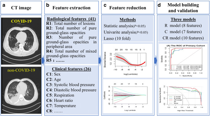
A consecutive cohort of 70 COVID-19 and 66 non-COVID-19 pneumonia patients were retrospectively recruited from five institutions. Patients were divided into primary (n = 98) and validation (n = 38) cohorts. The chi-square test, Student's t test, and Kruskal-Wallis H test were performed, comparing 1745 lesions and 67 features in the two groups. Three models were constructed using radiological semantic and clinical features through multivariate logistic regression. Diagnostic efficacies of developed models were quantified by receiver operating characteristic curve. Clinical usage was evaluated by decision curve analysis and nomogram. RESULTS: 
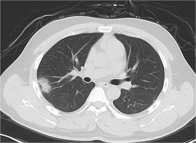
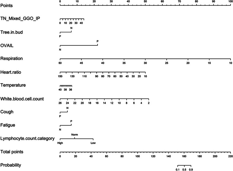
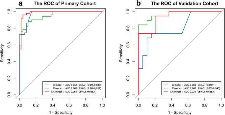
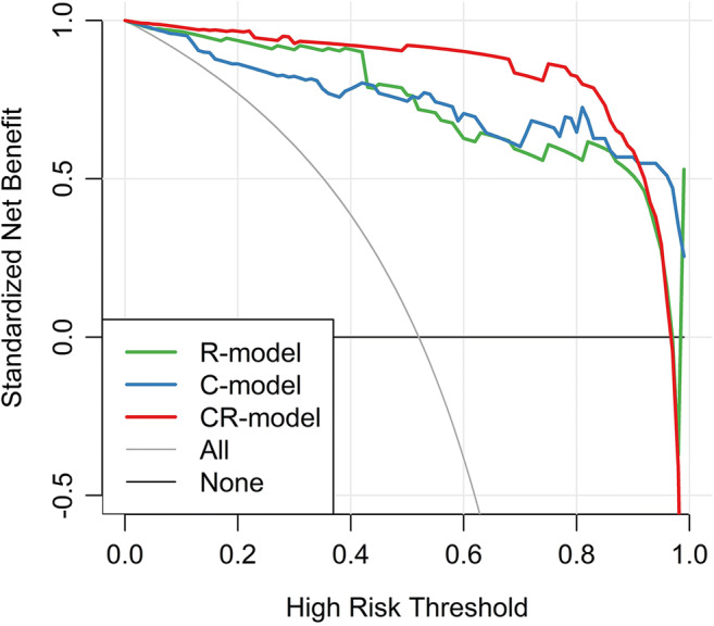
Eighteen radiological semantic features and seventeen clinical features were identified to be significantly different. Besides ground-glass opacities (p = 0.032) and consolidation (p = 0.001) in the lung periphery, the lesion size (1-3 cm) is also significant for the diagnosis of COVID-19 (p = 0.027). Lung score presents no significant difference (p = 0.417). Three diagnostic models achieved an area under the curve value as high as 0.986 (95% CI 0.9661.000). The clinical and radiological semantic models provided a better diagnostic performance and more considerable net benefits. CONCLUSIONS: Based on CT imaging and clinical manifestations alone, the pneumonia patients with and without COVID-19 can be distinguished. A model composed of radiological semantic and clinical features has an excellent performance for the diagnosis of COVID-19. KEY POINTS: • Based on CT imaging and clinical manifestations alone, the pneumonia patients with and without COVID-19 can be distinguished. • A diagnostic model for COVID-19 was developed and validated using radiological semantic and clinical features, which had an area under the curve value of 0.986 (95% CI 0.9661.000) and 0.936 (95% CI 0.866~1.000) in the primary and validation cohorts, respectively.

摘要

目的:在疫情期间,快速准确地诊断 2019 年冠状病毒病(COVID-19)至关重要。我们旨在确定患有 COVID-19 和非 COVID-19 肺炎患者的 CT 影像学和临床表现之间的差异,并开发和验证一种仅基于放射语义和临床特征的 COVID-19 诊断模型。

方法:回顾性纳入来自五家机构的 70 例 COVID-19 和 66 例非 COVID-19 肺炎患者的连续队列。患者分为主要队列(n=98)和验证队列(n=38)。采用卡方检验、学生 t 检验和 Kruskal-Wallis H 检验比较两组 1745 个病变和 67 个特征。通过多元逻辑回归利用放射语义和临床特征构建三个模型。通过受试者工作特征曲线量化所开发模型的诊断效能。通过决策曲线分析和列线图评估临床应用。

结果:确定了 18 个放射语义特征和 17 个临床特征存在显著差异。除了肺外周的磨玻璃影(p=0.032)和实变(p=0.001)外,病变大小(1-3cm)也对 COVID-19 的诊断具有显著意义(p=0.027)。肺评分无显著差异(p=0.417)。三个诊断模型的曲线下面积值高达 0.986(95%CI 0.966~1.000)。临床和放射语义模型提供了更好的诊断性能和更可观的净收益。

结论:仅基于 CT 影像学和临床表现,就可以区分患有 COVID-19 和非 COVID-19 的肺炎患者。由放射语义和临床特征组成的模型在 COVID-19 的诊断中具有出色的性能。

https://cdn.ncbi.nlm.nih.gov/pmc/blobs/1c2d/7431393/0397f3c4271b/330_2020_6829_Fig5_HTML.jpg
https://cdn.ncbi.nlm.nih.gov/pmc/blobs/1c2d/7431393/00b4e20cfbf6/330_2020_6829_Fig1_HTML.jpg
https://cdn.ncbi.nlm.nih.gov/pmc/blobs/1c2d/7431393/f1392c641aa8/330_2020_6829_Fig2_HTML.jpg
https://cdn.ncbi.nlm.nih.gov/pmc/blobs/1c2d/7431393/2d1edf3e7009/330_2020_6829_Fig3_HTML.jpg
https://cdn.ncbi.nlm.nih.gov/pmc/blobs/1c2d/7431393/a93e8c08bee6/330_2020_6829_Fig4_HTML.jpg
https://cdn.ncbi.nlm.nih.gov/pmc/blobs/1c2d/7431393/0397f3c4271b/330_2020_6829_Fig5_HTML.jpg

相似文献

[1]
A diagnostic model for coronavirus disease 2019 (COVID-19) based on radiological semantic and clinical features: a multi-center study.

Eur Radiol. 2020-4-16

[2]
Radiomics nomogram for the prediction of 2019 novel coronavirus pneumonia caused by SARS-CoV-2.

Eur Radiol. 2020-7-3

[3]
Analysis of clinical features and imaging signs of COVID-19 with the assistance of artificial intelligence.

Eur Rev Med Pharmacol Sci. 2020-8

[4]
Imaging and clinical features of patients with 2019 novel coronavirus SARS-CoV-2.

Eur J Nucl Med Mol Imaging. 2020-2-28

[5]
Development and Validation of a Prognostic Nomogram Based on Clinical and CT Features for Adverse Outcome Prediction in Patients with COVID-19.

Korean J Radiol. 2020-8

[6]
End-to-end automatic differentiation of the coronavirus disease 2019 (COVID-19) from viral pneumonia based on chest CT.

Eur J Nucl Med Mol Imaging. 2020-6-22

[7]
Nomogram to identify severe coronavirus disease 2019 (COVID-19) based on initial clinical and CT characteristics: a multi-center study.

BMC Med Imaging. 2020-10-2

[8]
Artificial Intelligence Augmentation of Radiologist Performance in Distinguishing COVID-19 from Pneumonia of Other Origin at Chest CT.

Radiology. 2020-4-27

[9]
Using Artificial Intelligence to Detect COVID-19 and Community-acquired Pneumonia Based on Pulmonary CT: Evaluation of the Diagnostic Accuracy.

Radiology. 2020-3-19

[10]
The Performance of Chest CT in Evaluating the Clinical Severity of COVID-19 Pneumonia: Identifying Critical Cases Based on CT Characteristics.

Invest Radiol. 2020-7

引用本文的文献

[1]
Guidance for clinical practice using emergency and point-of-care ultrasonography.

Acute Med Surg. 2024-6-26

[2]
Investigating the diagnostic and prognostic value of anti-SARS-CoV-2 Spike IgG/IgM ELISA tests in patients infected with coronavirus Delta variant.

Infez Med. 2024-3-1

[3]
Investigating of the role of CT scan for cancer patients during the first wave of COVID-19 pandemic.

Res Diagn Interv Imaging. 2022-3

[4]
A lightweight CORONA-NET for COVID-19 detection in X-ray images.

Expert Syst Appl. 2023-9-1

[5]
Developing medical imaging AI for emerging infectious diseases.

Nat Commun. 2022-11-18

[6]
Diagnostic models for fever of unknown origin based on F-FDG PET/CT: a prospective study in China.

EJNMMI Res. 2022-10-28

[7]
Novel chaotic oppositional fruit fly optimization algorithm for feature selection applied on COVID 19 patients' health prediction.

PLoS One. 2022

[8]
Development of Clinical Risk Scores for Detection of COVID-19 in Suspected Patients During a Local Outbreak in China: A Retrospective Cohort Study.

Int J Public Health. 2022

[9]
Cellular Imaging Analysis Algorithm-Based Assessment and Prediction of Disease in Patients with Acute Lung Injury.

Contrast Media Mol Imaging. 2022

[10]
Epidemiological challenges in pandemic coronavirus disease (COVID-19): Role of artificial intelligence.

Wiley Interdiscip Rev Data Min Knowl Discov. 2022

本文引用的文献

[1]
The Many Faces of COVID-19: Spectrum of Imaging Manifestations.

Radiol Cardiothorac Imaging. 2020-2-14

[2]
Imaging Profile of the COVID-19 Infection: Radiologic Findings and Literature Review.

Radiol Cardiothorac Imaging. 2020-2-13

[3]
Longitudinal CT Findings in COVID-19 Pneumonia: Case Presenting Organizing Pneumonia Pattern.

Radiol Cardiothorac Imaging. 2020-2-14

[4]
Radiological findings from 81 patients with COVID-19 pneumonia in Wuhan, China: a descriptive study.

Lancet Infect Dis. 2020-2-24

[5]
Correlation of Chest CT and RT-PCR Testing for Coronavirus Disease 2019 (COVID-19) in China: A Report of 1014 Cases.

Radiology. 2020-2-26

[6]
Sensitivity of Chest CT for COVID-19: Comparison to RT-PCR.

Radiology. 2020-8

[7]
Outbreak of novel coronavirus (COVID-19): What is the role of radiologists?

Eur Radiol. 2020-2-18

[8]
Initial CT findings and temporal changes in patients with the novel coronavirus pneumonia (2019-nCoV): a study of 63 patients in Wuhan, China.

Eur Radiol. 2020-2-13

[9]
Chest CT for Typical Coronavirus Disease 2019 (COVID-19) Pneumonia: Relationship to Negative RT-PCR Testing.

Radiology. 2020-2-12

[10]
Imaging changes in patients with 2019-nCov.

Eur Radiol. 2020-7

文献AI研究员

20分钟写一篇综述,助力文献阅读效率提升50倍

立即体验

用中文搜PubMed

大模型驱动的PubMed中文搜索引擎

马上搜索

推荐工具

医学文档翻译智能文献检索