• 文献检索
  • 文档翻译
  • 深度研究
  • 学术资讯
  • Suppr Zotero 插件Zotero 插件
  • 邀请有礼
  • 套餐&价格
  • 历史记录
应用&插件
Suppr Zotero 插件Zotero 插件浏览器插件Mac 客户端Windows 客户端微信小程序
定价
高级版会员购买积分包购买API积分包
服务
文献检索文档翻译深度研究API 文档MCP 服务
关于我们
关于 Suppr公司介绍联系我们用户协议隐私条款
关注我们

Suppr 超能文献

核心技术专利:CN118964589B侵权必究
粤ICP备2023148730 号-1Suppr @ 2026

文献检索

告别复杂PubMed语法,用中文像聊天一样搜索,搜遍4000万医学文献。AI智能推荐,让科研检索更轻松。

立即免费搜索

文件翻译

保留排版,准确专业,支持PDF/Word/PPT等文件格式,支持 12+语言互译。

免费翻译文档

深度研究

AI帮你快速写综述,25分钟生成高质量综述,智能提取关键信息,辅助科研写作。

立即免费体验

新型冠状病毒肺炎感染住院患者的胸部影像学——病例系列

Chest Imaging in Patients Hospitalized With COVID-19 Infection - A Case Series.

作者信息

Bhat Roopa, Hamid Aws, Kunin Jeffrey R, Saboo Sachin S, Batra Kiran, Baruah Dhiraj, Bhat Ambarish P

机构信息

Department of Radiology, University of Missouri, Columbia, MO.

Department of Radiology, University of Texas Health Science Center, San Antonio, TX..

出版信息

Curr Probl Diagn Radiol. 2020 Jul-Aug;49(4):294-301. doi: 10.1067/j.cpradiol.2020.04.001. Epub 2020 Apr 11.

DOI:10.1067/j.cpradiol.2020.04.001
PMID:32305134
原文链接:https://pmc.ncbi.nlm.nih.gov/articles/PMC7151480/
Abstract

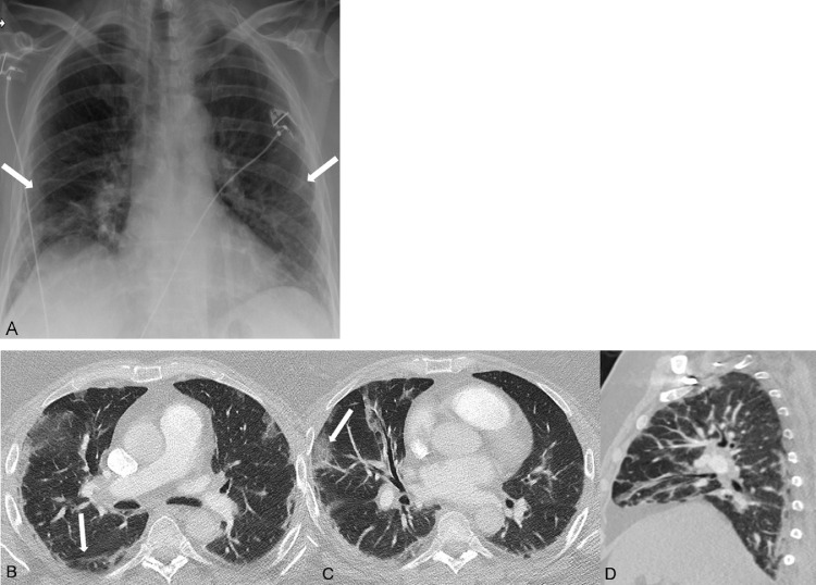
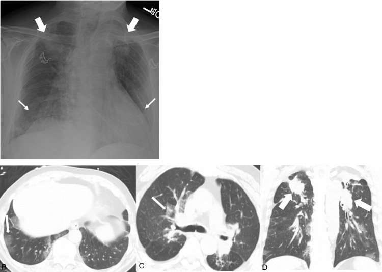
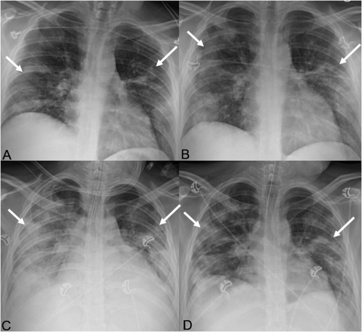
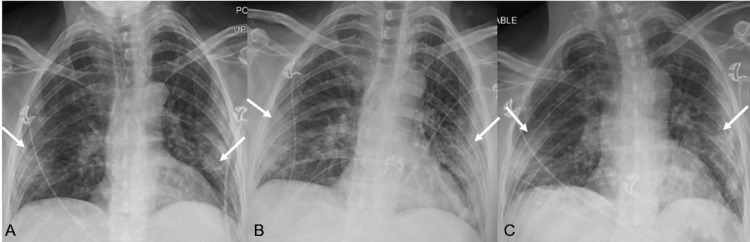
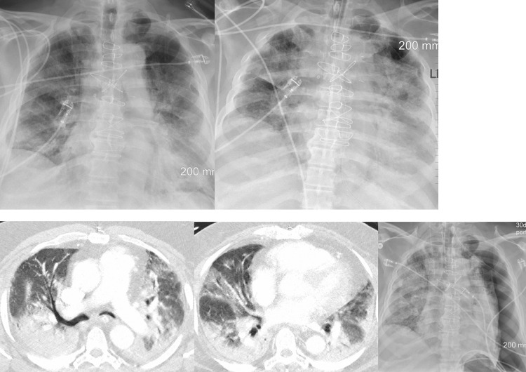
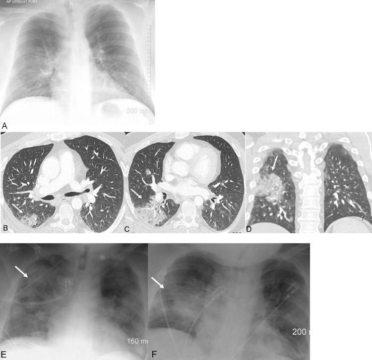
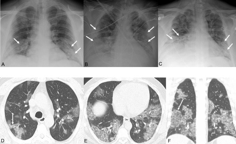
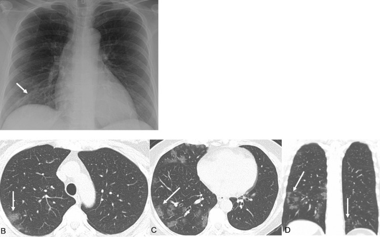
COVID-19 (Corona Virus Disease-19) is a zoonotic illness first reported in the city of Wuhan, China in December 2019, and is now officially a global pandemic as declared by the World Health Organization. The infection is caused by severe acute respiratory syndrome coronavirus 2 (SARS-CoV-2). COVID-19 infected patients can be asymptomatic carriers or present with mild-to-severe respiratory symptoms. Imaging, including computed tomography is not recommended to screen/diagnose COVID-19 infections, but plays an important role in management of these patients, and to rule out alternative diagnoses or coexistent diseases. In our multicenter case series, we outline the clinical presentations and illustrate the most common imaging manifestations in patients hospitalized with COVID-19.

摘要

2019冠状病毒病(COVID-19)是一种人畜共患病,于2019年12月在中国武汉市首次报告,目前世界卫生组织已宣布其为全球大流行疾病。该感染由严重急性呼吸综合征冠状病毒2(SARS-CoV-2)引起。COVID-19感染患者可能是无症状携带者,也可能出现轻至重度呼吸道症状。不建议使用包括计算机断层扫描在内的影像学检查来筛查/诊断COVID-19感染,但它在这些患者的管理中起着重要作用,并且有助于排除其他诊断或并存疾病。在我们的多中心病例系列中,我们概述了临床表现,并展示了COVID-19住院患者最常见的影像学表现。

https://cdn.ncbi.nlm.nih.gov/pmc/blobs/ed4c/7151480/66c9802dc886/gr8_lrg.jpg
https://cdn.ncbi.nlm.nih.gov/pmc/blobs/ed4c/7151480/d1d5518257d1/gr1_lrg.jpg
https://cdn.ncbi.nlm.nih.gov/pmc/blobs/ed4c/7151480/c1644c1662fe/gr2_lrg.jpg
https://cdn.ncbi.nlm.nih.gov/pmc/blobs/ed4c/7151480/42958b465bbc/gr3_lrg.jpg
https://cdn.ncbi.nlm.nih.gov/pmc/blobs/ed4c/7151480/3c864e5a339e/gr4_lrg.jpg
https://cdn.ncbi.nlm.nih.gov/pmc/blobs/ed4c/7151480/95daae46d2bd/gr5_lrg.jpg
https://cdn.ncbi.nlm.nih.gov/pmc/blobs/ed4c/7151480/395c997bfc09/gr6_lrg.jpg
https://cdn.ncbi.nlm.nih.gov/pmc/blobs/ed4c/7151480/97fc8776b7e1/gr7_lrg.jpg
https://cdn.ncbi.nlm.nih.gov/pmc/blobs/ed4c/7151480/66c9802dc886/gr8_lrg.jpg

相似文献

1
Chest Imaging in Patients Hospitalized With COVID-19 Infection - A Case Series.新型冠状病毒肺炎感染住院患者的胸部影像学——病例系列
Curr Probl Diagn Radiol. 2020 Jul-Aug;49(4):294-301. doi: 10.1067/j.cpradiol.2020.04.001. Epub 2020 Apr 11.
2
A case series of chest imaging manifestation of COVID-19.新型冠状病毒肺炎胸部影像学表现的病例系列
Radiography (Lond). 2020 Nov;26(4):e319-e321. doi: 10.1016/j.radi.2020.05.003. Epub 2020 May 14.
3
Thoracic imaging of coronavirus disease 2019 (COVID-19) in children: a series of 91 cases.儿童 2019 冠状病毒病(COVID-19)的胸部影像学表现:系列病例 91 例。
Pediatr Radiol. 2020 Sep;50(10):1354-1368. doi: 10.1007/s00247-020-04747-5. Epub 2020 Aug 4.
4
Thoracic imaging tests for the diagnosis of COVID-19.用于诊断新型冠状病毒肺炎的胸部影像学检查
Cochrane Database Syst Rev. 2020 Sep 30;9:CD013639. doi: 10.1002/14651858.CD013639.pub2.
5
Imaging findings in COVID-19 pneumonia.新型冠状病毒肺炎的影像学表现。
Clinics (Sao Paulo). 2020 Jun 22;75:e2027. doi: 10.6061/clinics/2020/e2027. eCollection 2020.
6
Initial chest CT findings in COVID-19: correlation with clinical features.COVID-19 的初始胸部 CT 表现:与临床特征的相关性。
J Zhejiang Univ Sci B. 2020;21(8):668-672. doi: 10.1631/jzus.B2000133.
7
Computed Tomography Manifestations of 5 Cases of the Novel Coronavirus Disease 2019 (COVID-19) Pneumonia From Patients Outside Wuhan.来自武汉以外地区的 5 例新型冠状病毒病(COVID-19)肺炎患者的计算机断层扫描表现。
J Thorac Imaging. 2020 May;35(3):W90-W93. doi: 10.1097/RTI.0000000000000508.
8
Respiratory Support in Severely or Critically Ill ICU Patients With COVID-19 in Wuhan, China.中国武汉 COVID-19 重症或危重症 ICU 患者的呼吸支持。
Curr Med Sci. 2020 Aug;40(4):636-641. doi: 10.1007/s11596-020-2227-8. Epub 2020 Aug 29.
9
Clinical characteristics of COVID-19 in 104 people with SARS-CoV-2 infection on the Diamond Princess cruise ship: a retrospective analysis.《104 名“钻石公主”号邮轮上 SARS-CoV-2 感染患者的 COVID-19 临床特征:一项回顾性分析》。
Lancet Infect Dis. 2020 Sep;20(9):1043-1050. doi: 10.1016/S1473-3099(20)30482-5. Epub 2020 Jun 12.
10
Computed tomography of covid-19 pneumonia.新型冠状病毒肺炎的计算机断层扫描
BMJ. 2020 Jul 9;370:m1807. doi: 10.1136/bmj.m1807.

引用本文的文献

1
Predicting the COVID-19 Patients Status Using Chest CT Scan Findings: A Risk Assessment Model Based on Decision Tree Analysis.基于决策树分析的胸部 CT 扫描结果预测 COVID-19 患者病情:风险评估模型。
Adv Exp Med Biol. 2023;1412:237-250. doi: 10.1007/978-3-031-28012-2_13.
2
An isolated spontaneous pneumomediastinum as a rare complication of COVID 19.孤立性自发性纵隔气肿作为 COVID-19 的罕见并发症。
Tunis Med. 2021;99(7):764-766.
3
Role of Chest Computed Tomography versus Real Time Reverse Transcription Polymerase Chain Reaction for Diagnosis of COVID-19: A Systematic Review and Meta-Analysis.

本文引用的文献

1
Radiological Society of North America Expert Consensus Document on Reporting Chest CT Findings Related to COVID-19: Endorsed by the Society of Thoracic Radiology, the American College of Radiology, and RSNA.北美放射学会关于报告与COVID-19相关的胸部CT检查结果的专家共识文件:得到了胸腔放射学会、美国放射学会和北美放射学会的认可。
Radiol Cardiothorac Imaging. 2020 Mar 25;2(2):e200152. doi: 10.1148/ryct.2020200152. eCollection 2020 Apr.
2
Frequency and Distribution of Chest Radiographic Findings in Patients Positive for COVID-19.COVID-19 阳性患者的胸部 X 线表现的频率和分布。
Radiology. 2020 Aug;296(2):E72-E78. doi: 10.1148/radiol.2020201160. Epub 2020 Mar 27.
3
胸部计算机断层扫描与实时逆转录聚合酶链反应在2019冠状病毒病诊断中的作用:一项系统评价和荟萃分析
Interdiscip Perspect Infect Dis. 2021 May 28;2021:8798575. doi: 10.1155/2021/8798575. eCollection 2021.
4
Highlighting COVID-19: What the imaging exams show about the disease.聚焦新冠疫情:影像学检查对该疾病的显示情况
World J Radiol. 2021 May 28;13(5):122-136. doi: 10.4329/wjr.v13.i5.122.
5
Computed tomography of the chest in patients with COVID-19: what do radiologists want to know?新型冠状病毒肺炎患者的胸部计算机断层扫描:放射科医生想了解什么?
Pol J Radiol. 2021 Feb 22;86:e122-e135. doi: 10.5114/pjr.2021.104049. eCollection 2021.
6
Delayed discharge is associated with higher complement C3 levels and a longer nucleic acid-negative conversion time in patients with COVID-19.延迟出院与 COVID-19 患者的补体 C3 水平升高和核酸转阴时间延长有关。
Sci Rep. 2021 Jan 13;11(1):1233. doi: 10.1038/s41598-021-81010-3.
7
Prevalence of COVID-19 Diagnostic Output with Chest Computed Tomography: A Systematic Review and Meta-Analysis.胸部计算机断层扫描对新型冠状病毒肺炎的诊断价值:一项系统评价与Meta分析
Diagnostics (Basel). 2020 Nov 28;10(12):1023. doi: 10.3390/diagnostics10121023.
8
Immunopathology, host-virus genome interactions, and effective vaccine development in SARS-CoV-2.严重急性呼吸综合征冠状病毒2(SARS-CoV-2)的免疫病理学、宿主-病毒基因组相互作用及有效疫苗开发
Comput Struct Biotechnol J. 2020;18:3774-3787. doi: 10.1016/j.csbj.2020.11.011. Epub 2020 Nov 20.
9
Review of X-ray and computed tomography scan findings with a promising role of point of care ultrasound in COVID-19 pandemic.X线及计算机断层扫描结果回顾,以及即时超声在COVID-19大流行中的潜在作用
World J Radiol. 2020 Sep 28;12(9):195-203. doi: 10.4329/wjr.v12.i9.195.
10
Using Artificial Intelligence for COVID-19 Chest X-ray Diagnosis.利用人工智能进行COVID-19胸部X光诊断。
Fed Pract. 2020 Sep;37(9):398-404. doi: 10.12788/fp.0045.
Clinical Features and Chest CT Manifestations of Coronavirus Disease 2019 (COVID-19) in a Single-Center Study in Shanghai, China.
中国上海单中心研究的 2019 年冠状病毒病(COVID-19)的临床特征和胸部 CT 表现。
AJR Am J Roentgenol. 2020 Jul;215(1):121-126. doi: 10.2214/AJR.20.22959. Epub 2020 Mar 14.
4
Coronavirus Disease 2019 (COVID-19): Role of Chest CT in Diagnosis and Management.新型冠状病毒病 2019(COVID-19):胸部 CT 在诊断和管理中的作用。
AJR Am J Roentgenol. 2020 Jun;214(6):1280-1286. doi: 10.2214/AJR.20.22954. Epub 2020 Mar 4.
5
Chest CT Findings in Patients With Coronavirus Disease 2019 and Its Relationship With Clinical Features.新型冠状病毒肺炎患者的胸部 CT 表现及其与临床特征的关系。
Invest Radiol. 2020 May;55(5):257-261. doi: 10.1097/RLI.0000000000000670.
6
An interactive web-based dashboard to track COVID-19 in real time.一个基于网络的交互式仪表盘,用于实时追踪新冠病毒。
Lancet Infect Dis. 2020 May;20(5):533-534. doi: 10.1016/S1473-3099(20)30120-1. Epub 2020 Feb 19.
7
Chest CT Findings in Coronavirus Disease-19 (COVID-19): Relationship to Duration of Infection.胸部计算机断层扫描在 2019 年冠状病毒病(COVID-19)中的表现:与感染持续时间的关系。
Radiology. 2020 Jun;295(3):200463. doi: 10.1148/radiol.2020200463. Epub 2020 Feb 20.
8
The novel Chinese coronavirus (2019-nCoV) infections: Challenges for fighting the storm.新型冠状病毒(2019 - nCoV)感染:抗击疫情面临的挑战
Eur J Clin Invest. 2020 Mar;50(3):e13209. doi: 10.1111/eci.13209. Epub 2020 Feb 5.
9
A Novel Coronavirus from Patients with Pneumonia in China, 2019.2019 年中国肺炎患者中的一种新型冠状病毒。
N Engl J Med. 2020 Feb 20;382(8):727-733. doi: 10.1056/NEJMoa2001017. Epub 2020 Jan 24.
10
The continuing 2019-nCoV epidemic threat of novel coronaviruses to global health - The latest 2019 novel coronavirus outbreak in Wuhan, China.新型冠状病毒持续的2019 - nCoV疫情对全球健康构成威胁——中国武汉最新的2019新型冠状病毒爆发。
Int J Infect Dis. 2020 Feb;91:264-266. doi: 10.1016/j.ijid.2020.01.009. Epub 2020 Jan 14.