• 文献检索
  • 文档翻译
  • 深度研究
  • 学术资讯
  • Suppr Zotero 插件Zotero 插件
  • 邀请有礼
  • 套餐&价格
  • 历史记录
应用&插件
Suppr Zotero 插件Zotero 插件浏览器插件Mac 客户端Windows 客户端微信小程序
定价
高级版会员购买积分包购买API积分包
服务
文献检索文档翻译深度研究API 文档MCP 服务
关于我们
关于 Suppr公司介绍联系我们用户协议隐私条款
关注我们

Suppr 超能文献

核心技术专利:CN118964589B侵权必究
粤ICP备2023148730 号-1Suppr @ 2026

文献检索

告别复杂PubMed语法,用中文像聊天一样搜索,搜遍4000万医学文献。AI智能推荐,让科研检索更轻松。

立即免费搜索

文件翻译

保留排版,准确专业,支持PDF/Word/PPT等文件格式,支持 12+语言互译。

免费翻译文档

深度研究

AI帮你快速写综述,25分钟生成高质量综述,智能提取关键信息,辅助科研写作。

立即免费体验

钙或磷共轭介孔二氧化硅纳米颗粒对牙本质小管封闭的疗效评估

Efficacy of Ca- or PO -conjugated mesoporous silica nanoparticles on dentinal tubule occlusion: an assessment.

作者信息

Yu Tianjia, Wang Yu, Cai Qiang, Wu Lin

机构信息

Department of Prosthodontics, School and Hospital of Stomatology, China Medical University, Liaoning Provincial Key Laboratory of Oral Diseases, Shenyang 110002, China.

School of Materials Science & Engineering, Tsinghua University, Beijing 100084, China.

出版信息

Ann Transl Med. 2020 Mar;8(5):173. doi: 10.21037/atm.2020.01.98.

DOI:10.21037/atm.2020.01.98
PMID:32309320
原文链接:https://pmc.ncbi.nlm.nih.gov/articles/PMC7154387/
Abstract

BACKGROUND

Maintaining a long-term biological effect of dental materials on dentinal tubule occlusion is one of the great technical challenges in dental clinics. In addition to physical treatment, chemical treatment to produce insoluble precipitates to seal dentinal tubules has been used. As dentin is mostly composed of calcium and phosphate complexes, in this study, we have developed a novel tubule-occluding material [Ca/PO @mesoporous silica nanoparticles (MSNs)] by separately conjugating either Ca or PO with MSNs.

METHODS

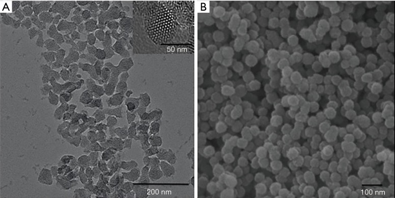
The shape and structure of the MSNs were examined using transmission electron microscopy (TEM) and scanning electron microscopy (SEM). The surface morphology and chemical compositions of Ca@MSNs/PO @MSNs and Ca/PO @MSNs were examined using SEM and X-ray fluorescence (XRF). The element distribution of Ca/PO @MSNs was detected using energy dispersive spectrometer (EDS). The sustained release ability of Ca@MSNs/PO @MSNs was detected using inductively coupled plasma atomic emission spectrometry (ICP-AES). The efficacy of Ca/PO @MSNs on dentinal tubule sealing was evaluated using SEM, and the results were analyzed by Image-Pro software to determine the best water-powder ratio. We also compared the sealing efficacy between Ca/PO @MSNs and NovaMin, which is currently used in clinics, under the simulated conditions of oral acidic corrosion and mechanical friction.

RESULTS

Ca/PO @MSNs are a new type of tubule-occluding material with sustained release properties. The ratio of Ca@MSNs: PO @MSNs: HO =0.015 g: 0.015 g: 150 µL exhibited an excellent sealing effect on dentinal tubules as well as resistance to oral acid corrosion and daily oral friction.

CONCLUSIONS

The novel dental material Ca/PO @MSNs demonstrates potential long-term effectiveness in sealing dentinal tubules and reducing dentin sensitivity, which is one of the most important problems in dental clinics.

摘要

背景

维持牙科材料对牙本质小管封闭的长期生物学效应是牙科临床中的重大技术挑战之一。除了物理处理外,还采用化学处理来产生不溶性沉淀物以封闭牙本质小管。由于牙本质主要由钙和磷酸盐复合物组成,在本研究中,我们通过将钙或磷分别与介孔二氧化硅纳米颗粒(MSNs)结合,开发了一种新型的小管封闭材料[Ca/PO@介孔二氧化硅纳米颗粒(MSNs)]。

方法

使用透射电子显微镜(TEM)和扫描电子显微镜(SEM)检查MSNs的形状和结构。使用SEM和X射线荧光(XRF)检查Ca@MSNs/PO@MSNs和Ca/PO@MSNs的表面形态和化学成分。使用能量色散光谱仪(EDS)检测Ca/PO@MSNs的元素分布。使用电感耦合等离子体原子发射光谱法(ICP-AES)检测Ca@MSNs/PO@MSNs的缓释能力。使用SEM评估Ca/PO@MSNs对牙本质小管封闭的效果,并通过Image-Pro软件分析结果以确定最佳水粉比。我们还在模拟口腔酸性腐蚀和机械摩擦的条件下,比较了Ca/PO@MSNs与目前临床使用的NovaMin之间的封闭效果。

结果

Ca/PO@MSNs是一种具有缓释特性的新型小管封闭材料。Ca@MSNs:PO@MSNs:HO的比例为0.015 g:0.015 g:150 μL时,对牙本质小管表现出优异的封闭效果以及对口腔酸腐蚀和日常口腔摩擦的抵抗力。

结论

新型牙科材料Ca/PO@MSNs在封闭牙本质小管和降低牙本质敏感性方面显示出潜在的长期有效性,牙本质敏感性是牙科临床中最重要的问题之一。

https://cdn.ncbi.nlm.nih.gov/pmc/blobs/440a/7154387/1717fa82ef5d/atm-08-05-173-f6.jpg
https://cdn.ncbi.nlm.nih.gov/pmc/blobs/440a/7154387/4d58d215d488/atm-08-05-173-f1.jpg
https://cdn.ncbi.nlm.nih.gov/pmc/blobs/440a/7154387/fa0ee35c71d0/atm-08-05-173-f2.jpg
https://cdn.ncbi.nlm.nih.gov/pmc/blobs/440a/7154387/a3c5a2733d69/atm-08-05-173-f3.jpg
https://cdn.ncbi.nlm.nih.gov/pmc/blobs/440a/7154387/41daf339acbb/atm-08-05-173-f4.jpg
https://cdn.ncbi.nlm.nih.gov/pmc/blobs/440a/7154387/9845a7531327/atm-08-05-173-f5.jpg
https://cdn.ncbi.nlm.nih.gov/pmc/blobs/440a/7154387/1717fa82ef5d/atm-08-05-173-f6.jpg

相似文献

1
Efficacy of Ca- or PO -conjugated mesoporous silica nanoparticles on dentinal tubule occlusion: an assessment.钙或磷共轭介孔二氧化硅纳米颗粒对牙本质小管封闭的疗效评估
Ann Transl Med. 2020 Mar;8(5):173. doi: 10.21037/atm.2020.01.98.
2
Effect of mesoporous silica nanoparticles on dentinal tubule occlusion: an in vitro study using SEM and image analysis.介孔二氧化硅纳米颗粒对牙本质小管封闭的影响:一项使用扫描电子显微镜和图像分析的体外研究
Dent Mater J. 2014;33(1):125-32. doi: 10.4012/dmj.2013-215.
3
Effect of different sizes of bioactive glass-coated mesoporous silica nanoparticles on dentinal tubule occlusion and mineralization.不同粒径的载有活性玻璃的介孔硅纳米颗粒对牙本质小管封闭和矿化的影响。
Clin Oral Investig. 2019 May;23(5):2129-2141. doi: 10.1007/s00784-018-2658-9. Epub 2018 Sep 30.
4
Application of electrophoretic deposition to occlude dentinal tubules in vitro.电泳沉积技术在体外封闭牙本质小管的应用。
J Dent. 2018 Apr;71:43-48. doi: 10.1016/j.jdent.2018.01.012. Epub 2018 Jan 31.
5
Comparison of in vitro dentinal tubule occluding efficacy of two different methods using a nano-scaled bioactive glass-containing desensitising agent.比较两种不同方法使用含纳米级生物活性玻璃脱敏剂对牙本质小管封闭效果的体外研究。
J Dent. 2017 May;60:63-69. doi: 10.1016/j.jdent.2017.03.001. Epub 2017 Mar 4.
6
Study on Occluding Dentinal Tubules with a Nanosilver-Loaded Silica System In Vitro.纳米银负载二氧化硅体系体外封闭牙本质小管的研究
ACS Omega. 2021 Jul 20;6(30):19596-19605. doi: 10.1021/acsomega.1c02123. eCollection 2021 Aug 3.
7
Dentin sealing and antibacterial effects of silver-doped bioactive glass/mesoporous silica nanocomposite: an in vitro study.银掺杂生物活性玻璃/介孔硅纳米复合材料对牙本质的密封和抗菌作用:一项体外研究。
Clin Oral Investig. 2019 Jan;23(1):253-266. doi: 10.1007/s00784-018-2432-z. Epub 2018 Apr 6.
8
Mesoporous Bioactive Glass Combined with Graphene Oxide Quantum Dot as a New Material for a New Treatment Option for Dentin Hypersensitivity.介孔生物活性玻璃与氧化石墨烯量子点复合作为牙本质过敏症新治疗方案的新型材料
Nanomaterials (Basel). 2020 Mar 27;10(4):621. doi: 10.3390/nano10040621.
9
Novel calcium encapsulated mesocellular siliceous foams for crystal growth in dentinal tubules.新型钙包埋介孔硅泡沫用于牙本质小管内晶体生长。
J Dent. 2019 Apr;83:61-66. doi: 10.1016/j.jdent.2019.02.007. Epub 2019 Feb 27.
10
Nano-CT characterization of dentinal tubule occlusion in SDF-treated dentin.SDF 处理牙本质中牙本质小管阻塞的纳米 CT 特征。
Sci Rep. 2023 Sep 23;13(1):15895. doi: 10.1038/s41598-023-42805-8.

引用本文的文献

1
Nano hydroxyapatite-silica with a core-shell structure for long-term management of dentin hypersensitivity.具有核壳结构的纳米羟基磷灰石-二氧化硅用于牙本质过敏的长期管理。
iScience. 2024 Nov 23;27(12):111474. doi: 10.1016/j.isci.2024.111474. eCollection 2024 Dec 20.
2
Nanotechnology in toothpaste: Fundamentals, trends, and safety.牙膏中的纳米技术:基础、趋势与安全性
Heliyon. 2024 Jan 20;10(3):e24949. doi: 10.1016/j.heliyon.2024.e24949. eCollection 2024 Feb 15.
3
Oyster shell-derived nano-hydroxyapatite and proanthocyanidin pretreatment on dentinal tubule occlusion and permeability before and after acid challenge-an in vitro study.

本文引用的文献

1
45S5 Bioglass paste is capable of protecting the enamel surrounding orthodontic brackets against erosive challenge.45S5生物活性玻璃糊剂能够保护正畸托槽周围的牙釉质免受侵蚀性刺激。
J Orthod Sci. 2019 Feb 20;8:5. doi: 10.4103/jos.JOS_93_18. eCollection 2019.
2
Evaluation of remineralization potential and mechanical properties of pit and fissure sealants fortified with nano-hydroxyapatite and nano-amorphous calcium phosphate fillers: An study.纳米羟基磷灰石和纳米无定形磷酸钙填料增强的窝沟封闭剂的再矿化潜力和力学性能评估:一项研究。
J Conserv Dent. 2018 Nov-Dec;21(6):681-690. doi: 10.4103/JCD.JCD_31_18.
3
Evaluation of biomimetically synthesized mesoporous silica nanoparticles as drug carriers: Structure, wettability, degradation, biocompatibility and brain distribution.
牡蛎壳衍生纳米羟基磷灰石和原花青素预处理对酸蚀前后牙本质小管闭塞和通透性的影响:一项体外研究。
J Mater Sci Mater Med. 2023 Apr 10;34(4):17. doi: 10.1007/s10856-023-06724-4.
4
Acidic Monetite Complex Paste with Bleaching Property for In-depth Occlusion of Dentinal Tubules.具有增白性能的酸性磷灰石复合糊剂用于牙本质小管的深度封闭。
Int J Nanomedicine. 2021 Jan 6;16:31-45. doi: 10.2147/IJN.S287393. eCollection 2021.
仿生合成介孔硅纳米粒子作为药物载体的评价:结构、润湿性、降解、生物相容性和脑分布。
Mater Sci Eng C Mater Biol Appl. 2019 Jan 1;94:453-464. doi: 10.1016/j.msec.2018.09.053. Epub 2018 Sep 22.
4
Preparation and Controllability of Mesoporous Silica Nanoparticles.介孔二氧化硅纳米粒子的制备与可控性
Enzymes. 2018;44:1-10. doi: 10.1016/bs.enz.2018.09.001. Epub 2018 Oct 15.
5
Effect of desensitizing toothpastes on dentin erosive wear and tubule occlusion. An in situ study.脱敏牙膏对牙本质侵蚀性磨损和牙本质小管封闭的影响:一项原位研究
Am J Dent. 2018 Aug;31(4):177-183.
6
Comparison between Fluoride and Nano-hydroxyapatite in Remineralizing Initial Enamel Lesion: An in vitro Study.氟化物与纳米羟基磷灰石对早期釉质龋再矿化作用的体外比较研究
J Contemp Dent Pract. 2018 Mar 1;19(3):306-312.
7
Application of electrophoretic deposition to occlude dentinal tubules in vitro.电泳沉积技术在体外封闭牙本质小管的应用。
J Dent. 2018 Apr;71:43-48. doi: 10.1016/j.jdent.2018.01.012. Epub 2018 Jan 31.
8
Development of Epigallocatechin-3-gallate-Encapsulated Nanohydroxyapatite/Mesoporous Silica for Therapeutic Management of Dentin Surface.没食子酸表没食子儿茶素酯-纳米羟基磷灰石/介孔硅的制备及其对牙本质表面治疗管理的应用
ACS Appl Mater Interfaces. 2017 Aug 9;9(31):25796-25807. doi: 10.1021/acsami.7b06597. Epub 2017 Jul 28.
9
Molecular Elucidation of Biological Response to Mesoporous Silica Nanoparticles in Vitro and in Vivo.体外和体内介孔二氧化硅纳米颗粒的生物学反应的分子阐明。
ACS Appl Mater Interfaces. 2017 Jul 12;9(27):22235-22251. doi: 10.1021/acsami.7b05359. Epub 2017 Jun 27.
10
Effect of Water-Cooled Nd:YAG Laser on Dentinal Tubule Occlusion In Vitro.水冷Nd:YAG激光对牙本质小管阻塞的体外影响
Photomed Laser Surg. 2017 Feb;35(2):98-104. doi: 10.1089/pho.2016.4169. Epub 2016 Nov 3.