Lemcke Heiko, Skorska Anna, Lang Cajetan Immanuel, Johann Lisa, David Robert
Department of Cardiac Surgery, Reference and Translation Center for Cardiac Stem Cell Therapy (RTC), Rostock University Medical Center, 18057 Rostock, Germany.
Faculty of Interdisciplinary Research, Department Life, Light & Matter, University Rostock, 18059 Rostock, Germany.
Int J Mol Sci. 2020 Apr 17;21(8):2819. doi: 10.3390/ijms21082819.
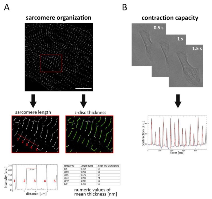
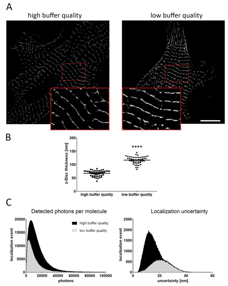
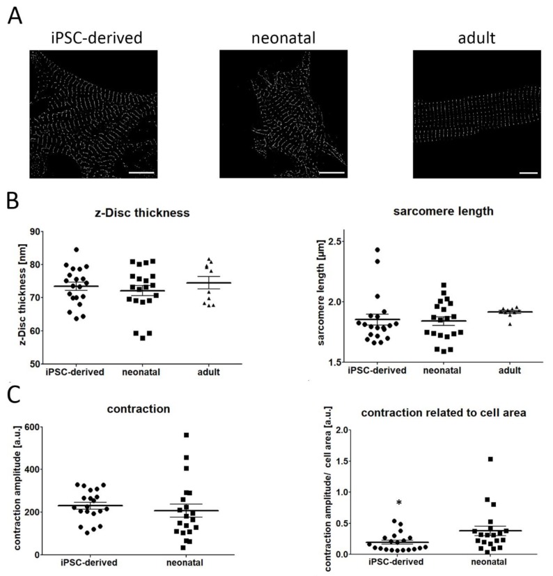
The maturation of iPSC-derived cardiomyocytes is still a critical point for their application in cardiovascular research as well as for their clinical use. Although multiple differentiation protocols have been established, researchers failed to generate fully mature cardiomyocytes in vitro possessing identical phenotype-related and functional properties as their native adult counterparts. Besides electrophysiological and metabolic changes, the establishment of a well structured sarcomere network is important for the development of a mature cardiac phenotype. Here, we present a super resolution-based approach to quantitatively evaluate the structural maturation of iPSC-derived cardiomyocytes. Fluorescence labelling of the α-actinin cytoskeleton and subsequent visualization by photoactivated localization microscopy allows the acquisition of highly resolved images for measuring sarcomere length and z-disc thickness. Our image analysis revealed that iPSC and neonatal cardiomyocyte share high similarity with respect to their sarcomere organization, however, contraction capacity was inferior in iPSC-derived cardiac cells, indicating an early maturation level. Moreover, we demonstrate that this imaging approach can be used as a tool to monitor cardiomyocyte integrity, helping to optimize iPSC differentiation as well as somatic cell direct-reprogramming strategies.
诱导多能干细胞衍生的心肌细胞的成熟,对于其在心血管研究中的应用以及临床应用而言,仍然是一个关键点。尽管已经建立了多种分化方案,但研究人员未能在体外生成具有与天然成年心肌细胞相同的表型相关和功能特性的完全成熟的心肌细胞。除了电生理和代谢变化外,构建结构良好的肌节网络对于成熟心脏表型的发展也很重要。在此,我们提出一种基于超分辨率的方法来定量评估诱导多能干细胞衍生的心肌细胞的结构成熟度。对α-辅肌动蛋白细胞骨架进行荧光标记,随后通过光激活定位显微镜进行可视化,从而能够获取用于测量肌节长度和Z盘厚度的高分辨率图像。我们的图像分析表明,诱导多能干细胞衍生的心肌细胞和新生心肌细胞在肌节组织方面具有高度相似性,然而,诱导多能干细胞衍生的心肌细胞的收缩能力较差,表明其处于早期成熟水平。此外,我们证明这种成像方法可作为监测心肌细胞完整性的工具,有助于优化诱导多能干细胞分化以及体细胞直接重编程策略。