Hernández Cristina, Porta Massimo, Bandello Francesco, Grauslund Jakob, Harding Simon P, Aldington Stephen J, Egan Catherine, Frydkjaer-Olsen Ulrik, García-Arumí José, Gibson Jonathan, Lang Gabriele E, Lattanzio Rosangela, Massin Pascale, Midena Edoardo, Ponsati Berta, Ribeiro Luísa, Scanlon Peter, Cunha-Vaz José, Simó Rafael
Diabetes and Metabolism Research Unit and CIBERDEM, Vall d'Hebron Research Institute, 08035 Barcelona, Spain.
Department of Medical Sciences, University of Turin, 10124 Turin, Italy.
J Clin Med. 2020 Apr 24;9(4):1233. doi: 10.3390/jcm9041233.
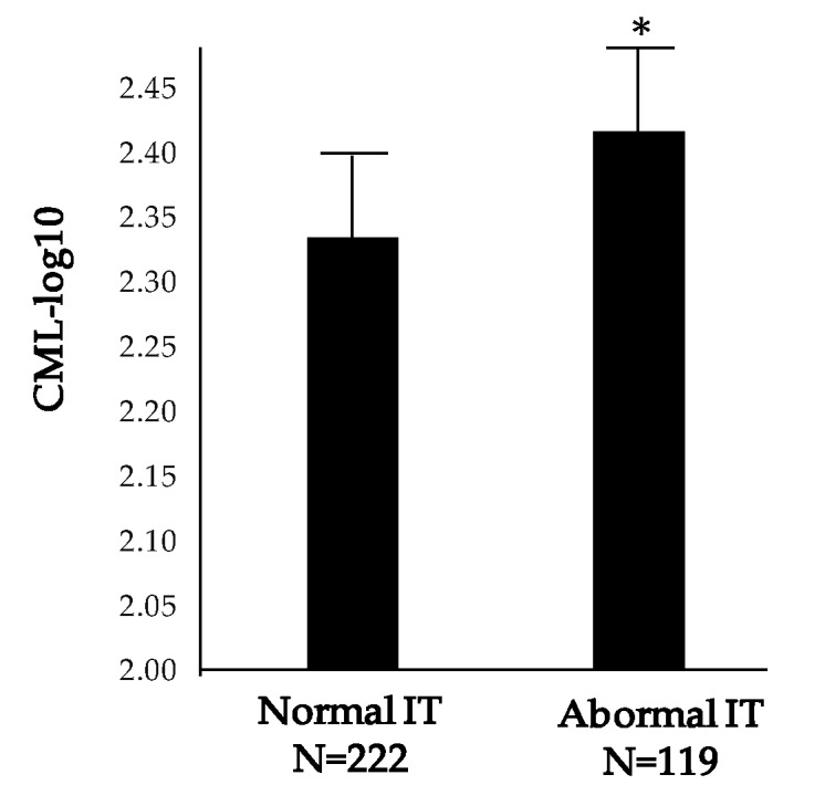
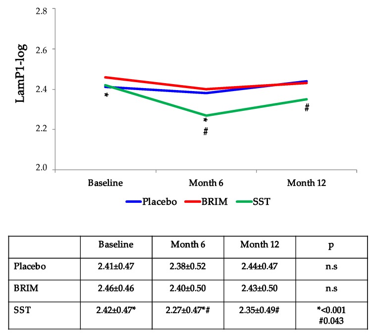
The main aim of this study was to evaluate the ability of serum biomarkers to predict the worsening of retinal neurodysfunction in subjects with type 2 diabetes. For this purpose, we measured selected molecules (N-epsilon-carboxy methyl lysine (CML), laminin P1 (Lam-P1), and asymmetric dimethylarginine (ADMA)) in the serum of 341 participants of the EUROCONDOR study at baseline, 24, and 48 weeks. Retinal neurodysfunction was assessed by measuring implicit time (IT) using multifocal electroretinography, and structural changes were examined by spectral domain-optical coherence tomography. The values of IT at baseline were directly correlated with baseline serum concentrations of CML ( = 0.135, = 0.013). Furthermore, in the placebo group, increase in CML concentration throughout follow-up correlated with the IT ( = 0.20; = 0.03). Baseline serum levels of CML also correlated with macular retinal thickness (RT) ( = 0.231; < 0.001). Baseline Lam-P1 levels correlated with the increase of the RT at the end of follow-up in the placebo group ( = 0.22; = 0.016). We provide evidence that CML may be a biomarker of both retinal neurodysfunction and RT, whereas Lam-P1 was associated with RT only. Therefore, circulating levels of these molecules could provide a complementary tool for monitoring the early changes of diabetic retinopathy (DR).
本研究的主要目的是评估血清生物标志物预测2型糖尿病患者视网膜神经功能障碍恶化的能力。为此,我们在EUROCONDOR研究的341名参与者的血清中,于基线、24周和48周时测量了选定的分子(N-ε-羧甲基赖氨酸(CML)、层粘连蛋白P1(Lam-P1)和不对称二甲基精氨酸(ADMA))。通过使用多焦视网膜电图测量隐时(IT)来评估视网膜神经功能障碍,并通过光谱域光学相干断层扫描检查结构变化。基线时的IT值与CML的基线血清浓度直接相关(= 0.135,= 0.013)。此外,在安慰剂组中,随访期间CML浓度的增加与IT相关(= 0.20;= 0.03)。CML的基线血清水平也与黄斑视网膜厚度(RT)相关(= 0.231;< 0.001)。安慰剂组中,基线Lam-P1水平与随访结束时RT的增加相关(= 0.22;= 0.016)。我们提供的证据表明,CML可能是视网膜神经功能障碍和RT的生物标志物,而Lam-P1仅与RT相关。因此,这些分子的循环水平可为监测糖尿病视网膜病变(DR)的早期变化提供一种补充工具。